Medical Ultrasound Imaging
Sunday, 19 May 2024
Info Sheets Out- side     | Ultrasound Database • 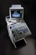 From Philips Medical Systems;
'The powerful system... The SD 260 is a powerful B/W ultrasound system that offers standard all major scan techniques: curved, linear and annular phased array. The user has a choice of no less than 12 probes and can connect up to three probes simultaneously, regardless of their technique. The SD 260 is perfectly suited for use in either a busy hospital or clinic.' This product is not available in the US, Australia, or New Zealand from Philips Medical Systems. Specifications for this system will be available soon. • 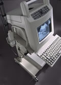 From Philips Medical Systems;
'A perfect portable system... The SD 100 is a system designed to travel. Weighing only 10.5 kg and battery or mains operated, the SD 100E is a full-featured ultrasound unit with a comprehensive software package, nine-inch monitor, digital output and built-in diskette for software updates and image storage. The SD 100E user can choose from seven linear and convex array dual frequency probes to perform many ultrasound examinations, quickly and reliably.' This product is not available in the US, Australia, or New Zealand from Philips Medical Systems. Specifications for this system will be available soon. •
The second generation ultrasound contrast agents (UCA/USCA) are both sufficiently small and stable to pass into the systemic circulation, and these contrast media enhance the Doppler signal in various arteries after intravenous injection. Second generation agents have a short live, the contrast effect is over in a few minutes.
•
Shear Waves are waves that travel perpendicular to the direction of the sound beam. During an ultrasound examination, shear waves are generated and transmitted into the body using the ultrasound probe. These waves travel through the tissue and their speed of propagation is influenced by the tissue's stiffness. Softer tissues allow the shear waves to travel faster, while stiffer tissues slow them down.
By analyzing the speed of shear waves, ultrasound systems can provide quantitative measurements of tissue stiffness, known as shear wave elastography. This technique is particularly useful in assessing the stiffness of organs like the liver, breast, thyroid, and muscles.
Shear wave ultrasound elastography has various applications in clinical practice. For example, it can help identify liver fibrosis, a condition characterized by excessive scarring of the liver tissue. By measuring the liver's stiffness using shear waves, clinicians can assess the severity of fibrosis without the need for invasive procedures like liver biopsies.
• View NEWS results for 'Shear Wave' (5). Further Reading: News & More:
•
Diagnostic ultrasound imaging has no known risks or long-term side effects. Discomfort to the patient is very rare if the sonogram is accurately performed by using appropriate frequencies and intensity ranges. However, the application of the ALARA principle is always recommended.
There are reports of low birth weight of babies after applying more than the recommended ultrasound examinations during pregnancy. Women who think they might be pregnant should raise this issue with the doctor before undergoing an abdominal ultrasound, to avoid any harm to the fetus in the early stages of development. Since ultrasound is energy, sensitive tissues like the reproductive organs could possibly sustain damage if vibrated to a high degree by too intense ultrasound waves. In diagnostic ultrasonic procedures, such damage would only result from improper use of the equipment. Possible ultrasound bioeffects:
•
•
Due to increasing of temperature, dissolved gases from microbubbles come out of the contrast solution.
The thermal effect is controlled by the displayed thermal index and the mechanical index indicates the risk of cavitation. An ultrasound gel is applied to obtain better contact between the transducer and the skin. This has the consistency of thick mineral oil and is not associated with skin irritation or allergy. Specific conditions for which ultrasound may be selected as a treatment may be attached with higher risks. See also Ultrasound Imaging Procedures, Fetal Ultrasound and Obstetric and Gynecologic Ultrasound. • View NEWS results for 'Side Effect' (7). | Share This Page Look Ups |
Medical-Ultrasound-Imaging.com
former US-TIP.com
Member of SoftWays' Medical Imaging Group - MR-TIP • Radiology TIP • Medical-Ultrasound-Imaging
Copyright © 2008 - 2024 SoftWays. All rights reserved.
Terms of Use | Privacy Policy | Advertise With Us
former US-TIP.com
Member of SoftWays' Medical Imaging Group - MR-TIP • Radiology TIP • Medical-Ultrasound-Imaging
Copyright © 2008 - 2024 SoftWays. All rights reserved.
Terms of Use | Privacy Policy | Advertise With Us
[last update: 2023-11-06 01:42:00]