Medical Ultrasound Imaging
Wednesday, 8 May 2024
Info Sheets Out- side     | 'Thyroid Ultrasound' Searchterm 'Thyroid Ultrasound' found in 13 articles 1 term [ • ] - 1 definition [• ] - 11 booleans [• ]Result Pages : • Thyroid Ultrasound
A thyroid ultrasound evaluates the size and shape of the thyroid gland and parathyroid glands. A thyroid ultrasound can show nodules, cysts, tumors, and an enlargement, but a sonogram cannot determine the function of the thyroid. Ultrasound guides the placement of the needle during a thyroid fine needle aspiration biopsy. See also Sonographic Features, Ultrasound Imaging Modes, Anechoic, Beam Width Artifact and Enhancement Artifact. •
Interventional ultrasound, also known as ultrasonography, encompasses a range of invasive or surgical procedures guided by ultrasound imaging. While its widest application lies in intravascular ultrasound imaging for measuring atherosclerotic plaque, it has proven valuable in various medical fields. In urology, ultrasound-guided interventions are employed for treatments like high intensity focused ultrasound (HIFU) in prostate conditions. The precise imaging provided by ultrasound aids in targeting the affected area and delivering therapeutic energy effectively. In intraabdominal conditions, endoscopic ultrasound is frequently utilized. This technique combines ultrasound imaging with an endoscope to visualize and evaluate structures within the gastrointestinal tract, allowing for precise diagnoses and targeted interventions. Ultrasound-guided procedures play a significant role in several medical specialties, including liver sonography, obstetric and gynecologic ultrasound, and thyroid ultrasound. These procedures involve interventions such as RF thermal ablation or biopsies, which are guided by real-time ultrasound imaging. For instance, in liver sonography, ultrasound guidance is crucial for performing biopsies or RF thermal ablation, a technique used to treat liver tumors by delivering localized heat to destroy the abnormal tissue. The real-time imaging allows for precise needle placement and monitoring during the procedure. In obstetric and gynecologic ultrasound, ultrasound-guided procedures, such as biopsies, can be performed to obtain tissue samples for diagnostic purposes. Additionally, ultrasound guidance is valuable during interventions like amniocentesis or fetal blood sampling, enabling accurate and safe procedures. Thyroid ultrasound procedures often involve ultrasound-guided fine-needle aspiration biopsy (FNAB), which allows for the sampling of thyroid nodules for cytological examination. The ultrasound image helps guide the needle into the targeted area, ensuring accurate sampling and minimizing potential complications. Overall, ultrasound-guided interventions provide minimally invasive and precise approaches to diagnosis and treatment. The real-time imaging capabilities of ultrasound contribute to enhanced accuracy, safety, and patient outcomes in procedures like biopsies, injections, and drainage. See also Transurethral Sonography, Endocavitary Echography, and B-Mode Acquisition and Targeting. • 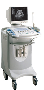
From SIUI Inc.;
Device Information and Specification
CONFIGURATION
Normal system, gray scale(256)
Linear and convex
PROBES STANDARD
2.5MHz ~ 11.0MHz, broad band, tri-frequency
IMAGING OPTIONS
Real zoom, max. Zoom * 4.0, multiplying power and position selectable
OPTIONAL PACKAGE
H*W*D m
1.30 * 0.52 * 0.77
WEIGHT
90kg (main unit)
POWER REQUIREMENT
AC 220V/110V, 50Hz/60Hz
POWER CONSUMPTION
0.45 KVA
•  From SIUI Inc.;
'Dedicated to ultrasound industry, Shantou Institute of Ultrasonic Instruments, Inc. (SIUI) has launched Apogee 3500, the Digital Color Doppler Ultrasound Imaging System. With latest imaging technologies, high-definition image quality and excellent practical functions, the Apogee 3500 offers optimal solutions for clinical ultrasonic examination.' 'The Apogee 3500 is available with many high-density, super broadband and multi-frequency probes, such as convex, micro-convex, linear, vaginal, rectal and phased array probes, which are widely applied for different clinical diagnoses, including abdomen (liver, kidney, gall-bladder, pancreas), gynecology (uterus, ovary), obstetrics (early pregnancy, basic OB, complete OB, multi gestation, fetal echo), cardiology (adult and pediatric cardiology), small parts (thyroid, galactophore, testicles, neonate), peripheral vascular and prostate.'
Device Information and Specification
CONFIGURATION
Normal system, color - gray scale(256)
Linear, convex and phased array
PROBES STANDARD
2.0 MHz ~ 12.0 MHz, broad band, tri-frequency
B-mode (B, 2B, 4B), M-mode, B/M-mode, real-time compound imaging, panoramic imaging, trapezoidal imaging (linear probes), spectrum Doppler (PWD and CWD), color Doppler flow imaging (CDFI), color power angio (CPA), tissue harmonic imaging (THI)
IMAGING OPTIONS
Real-time ZOOM, zoom rate and position selectable
OPTIONAL PACKAGE
H*W*D m
1.29 * 0.52 * 0.75
WEIGHT
110 kg
POWER REQUIREMENT
AC 220V/110V, 50Hz/60Hz
POWER CONSUMPTION
0.6 KVA
•  From SIUI Inc.;
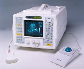
'The CTS-285 is designed for the diagnosis of liver, gallbladder, kidney, pancreas, thyroid, breast, uterus, bladder, ovary, etc. It is a portable versatile ultrasound scanner with convex and linear array scanning.'
Device Information and Specification
APPLICATIONS
See description above
CONFIGURATION
Portable, gray scale(256)
Linear and convex
PROBES STANDARD
1 * 2.5MHz ~ 5.0MHz trifrequency convex probe
2.5MHz to 10.0MHz, linear and convex, broad band, trifrequency
IMAGING OPTIONS
Multi zoom rate and depth shift
DATA PROCESSING
Pre-processing, correlation-processing, interpolation
POWER REQUIREMENT
AC 220V/110V, 50Hz/60Hz
POWER CONSUMPTION
0.06 KVA
Result Pages : | Share This Page Look Ups |
Medical-Ultrasound-Imaging.com
former US-TIP.com
Member of SoftWays' Medical Imaging Group - MR-TIP • Radiology TIP • Medical-Ultrasound-Imaging
Copyright © 2008 - 2024 SoftWays. All rights reserved.
Terms of Use | Privacy Policy | Advertise With Us
former US-TIP.com
Member of SoftWays' Medical Imaging Group - MR-TIP • Radiology TIP • Medical-Ultrasound-Imaging
Copyright © 2008 - 2024 SoftWays. All rights reserved.
Terms of Use | Privacy Policy | Advertise With Us
[last update: 2023-11-06 01:42:00]