Medical Ultrasound Imaging
Wednesday, 8 May 2024
Info Sheets Out- side     | 'Laparoscopic Ultrasound' Searchterm 'Laparoscopic Ultrasound' found in 7 articles 1 term [ • ] - 2 definitions [• ] - 4 booleans [• ]Result Pages : • Laparoscopic Ultrasound
(LUS) Diagnostic laparoscopy combined with laparoscopic ultrasound is used for staging tumors and to monitor surgical interventions like for example radiofrequency ablation or cryotherapy. Laparoscopic ultrasound provides direct contact imaging of organs with high frequency ultrasound. Laparoscopic ultrasound identifies and characterizes the tumor, guides the probe, and monitors the progression of the freezing or the thermal destruction. This procedure avoid unnecessary open surgery and improves selection of patients for tumor resection e.g., in liver and pancreas. Challenges of LUS are limitations of the intraoperative acoustic windows and the possible movement of the probe and that standard orientation techniques are difficult to apply with laparoscopic instruments, resulting in images from oblique planes. 3D ultrasound or special navigation systems may be helpful. See also Ultrasound Therapy. • View NEWS results for 'Laparoscopic Ultrasound' (1). •
The usual applications of endocavitary echography (also called internal echography / endoscopic ultrasound (EUS)) are examinations of the pelvic organs through internally introduced probes, which give a more precise and correct image. Transrectal ultrasound is a well established method for rectal or prostate carcinoma assessment. A transvaginal echography uses a small transducer that is inserted directly into the vagina. Used are high-frequency (10-12 MHz) for superficial organs, endocavitary echography, and intraoperative laparoscopic ultrasound. A sterile cover is slipped over the probe, which is then covered with lubricating ultrasound gel and placed in the cavitary (see Equipment Preparation). See also Endoscopic Ultrasound, Prostate Ultrasound, Interventional Ultrasound, Transurethral Sonography, Vaginal Probe, Rectal Probe. •
Most usual ultrasound machines are 2D real-time systems. This types of ultrasound scanners allow to assess both motion and anatomy, including the motion of heart valves, the movement of intestines and lungs and also to guide interventions, like for example a biopsy or a laparoscopic ultrasound. A standard real-time scanner consists of a mobile console with the monitor on the top and rows of small containers at the bottom to accommodate a variety of scanner probes. The linear, curved or phased array transducers are usually equipped with multiple crystals or in some cases with a moving crystal. A real-time scanner may be e.g., a mechanical scanner or electronic array scanner. See also Musculoskeletal and Joint Ultrasound. Further Reading: News & More: • 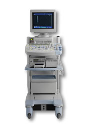 From Hitachi Medical Corporation (HMC), sales, marketing and service in the US by Hitachi Medical Systems America Inc.
The HI VISION™ 5500 - EUB-5500 fully digital ultrasound system delivers the latest generation of signal processing technology, sophisticated transducer design, and a host of features and options for advanced imaging capabilities across a wide range of clinical situations. This system is compatible with all Pentax ultrasound endoscopes.
Device Information and Specification
APPLICATIONS
Abdominal, brachytherapy/cryotherapy, breast, cardiac, dedicated biopsy, endoscopic, intraoperative, laparoscopic, musculoskeletal, OB/GYN, pediatric, small parts, urologic, vascular
CONFIGURATION
Compact system
Five frequency (except mini-probes)
Linear, convex, radial, miniradial/miniprobe, biplane, phased array, echoendoscope longitudinal, echoendoscope radial
3 modes of dynamic tissue harmonic imaging (dTHI), pulsed wave Doppler, continuous wave Doppler, color flow imaging, power Doppler, directional power Doppler, color flow angiography, real-time Doppler measurements
IMAGING OPTIONS
3RD generation color artifact suppression
OPTIONAL PACKAGE
STORAGE, CONNECTIVITY, OS
Patient and image database management system, HDD, FDD, MOD, CD-ROM, Network, DICOM 3.0, Windows XP
DATA PROCESSING
H*W*D m (inch.)
1.40 x 0.51 x 0.79 (55 x 20 x 31)
WEIGHT
130 kg (286 lbs.)
POWER CONSUMPTION
1.2kVA
ENVIRONMENTAL IMPACT
4096 btu/hr heat output
•  From Hitachi Medical Corporation (HMC), sales, marketing and service in the US by Hitachi Medical Systems America Inc.;
Powerful, flexible, and fast, the HI VISION™ 8500 - EUB-8500 diagnostic ultrasound scanner combines leading edge technologies with user-oriented operation for exceptional imaging and functionality. Available exclusively on the 8500, SonoElastography provides a new perspective on the physical properties of tumors and masses by determining and displaying the relative stiffness of tissue. Device Information and Specification
APPLICATIONS
Abdominal, brachytherapy/cryotherapy, breast, cardiac, dedicated biopsy, endoscopic, intraoperative, laparoscopic, musculoskeletal, OB/GYN, pediatric, small parts, urologic, vascular
CONFIGURATION
Compact system
RANGE OF PROBE TYPE
Linear, convex, radial, biplane, phased array, echoendoscope longitudinal, echoendoscope radial
PROBE FREQUENCIES
Linear: 5.0-13 MHz, convex: 2.5-7.5 MHz, phased:
2.0-7.5 MHz, sector: 2.0-7.5 MHz
4 Modes of dynamic tissue harmonic imaging (dTHI), pulsed wave Doppler, continuous wave Doppler, color flow imaging, power Doppler, directional power Doppler, color flow angiography, real-time Doppler measurements, quantitative tissue Doppler
IMAGING OPTIONS
HI COMPOUND imaging,
HI RES adaptive imaging, wideband pulse inversion imaging (WPI), Raw Data Freeze
OPTIONAL PACKAGE
IMAGING ENHANCEMENTS
3RD generation color artifact suppression
STORAGE, CONNECTIVITY, OS
Patient and image database management system, HDD, FDD, MOD, CD-ROM, Network, DICOM 3.0, Windows XP
DATA PROCESSING
Octal beam processing, 12 bit Gigasampling A/D for precise signal reproduction
H*W*D m (inch.)
1.50 * 0.56 * 1.02 (59 x 22 x 40)
WEIGHT
159 kg (351 lbs.)
POWER CONSUMPTION
1.5kVA
Result Pages : | Share This Page Look Ups |
Medical-Ultrasound-Imaging.com
former US-TIP.com
Member of SoftWays' Medical Imaging Group - MR-TIP • Radiology TIP • Medical-Ultrasound-Imaging
Copyright © 2008 - 2024 SoftWays. All rights reserved.
Terms of Use | Privacy Policy | Advertise With Us
former US-TIP.com
Member of SoftWays' Medical Imaging Group - MR-TIP • Radiology TIP • Medical-Ultrasound-Imaging
Copyright © 2008 - 2024 SoftWays. All rights reserved.
Terms of Use | Privacy Policy | Advertise With Us
[last update: 2023-11-06 01:42:00]