Medical Ultrasound Imaging
Wednesday, 8 May 2024
Info Sheets Out- side     | 'Pulsed Wave Doppler' Searchterm 'Pulsed Wave Doppler' found in 27 articles 1 term [ • ] - 18 definitions [• ] - 8 booleans [• ]Result Pages : • Pulsed Wave Doppler
(PWD) Pulsed wave (PW) Doppler is a Doppler ultrasound mode that evaluates blood flow velocities in a range specific area along the length of the sound beam. Measured are changes in received frequency due to relative motion (flow) between a sound source (transducer) and sound receiver (transducer). PW Doppler produces an audible signal as well as a graphical representation of flow. The Doppler shift produced by moving blood flow is calculated by the ultrasound system. See also Amplitude Indicator, Pulsed Ultrasound. Further Reading: News & More: • 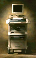 From SIUI Inc.;
'The Apogee 800Plus ultrasound system offers a new level of high quality in image. With broad clinical application, the system delivers ease of use for cardiac and vascular exams.' 'Due to the exceptional image quality, the sensitivity of color Doppler and many other advanced features, the Apogee 800Plus offers complete diagnosis capability for your entire patient population, including abdominal, obstetrics, gynecology, adult cardiology, pediatric cardiology, peripheral vascular, small parts, breast, prostate, transcranial, etc.'
Device Information and Specification
APPLICATIONS
Linear, curved, phased and symmetric phased array
2D, M-mode, Pulsed wave Doppler, Color Power Angio Imaging, Color 2D, Color M-mode, High PRF Doppler
H*W*D m
1.46 * 0.69 * 0.87
WEIGHT
140 kg (without peripherals)
POWER REQUIREMENT
115Vac, (V~), @ 50 to 60 Hz, 1.2kVA
230Vac, (V~), @ 50 to 60 Hz, 1.2kVA POWER CONSUMPTION
800 Watts (with optional OEM'S: 1200 Watts max)
•
Doppler techniques are dependent on the transducers used. The transducer operating in continuous wave mode utilizes one half of the elements and is continuously sending sound energy while the other half is continuously receiving the reflected signals. If the transducer is being used in a pulsed wave mode, the whole transducer is used to send and then receive the returning signals. Pulsed wave techniques allow the accurate measurement of blood flow at a specific area in the heart and the detection of both velocity and direction. Measurement is performed by timing the reception of the returning signals giving a view of flows at specific depths. The region where flow velocities are measured is called the sample volume. Errors in the accuracy of the information arise if the velocities exceed a certain speed. The highest velocity accurately measured is called the Nyquist limit.
•
Continuous Wave Doppler
Used for accurate measurement of high Velocity flow. A disadvantage is the poor range of resolution.
•
Pulsed Wave Doppler
Used for the measurement of velocities at a specific location with a good range of resolution. A disadvantage is the imprecise measuring of high velocities. See also Doppler Velocity Signal and Doppler Effect. Further Reading: Basics:
News & More:
•
Doppler ultrasound is a medical imaging technique for calculating the relative velocity between two points by measuring the frequency shift of a sound wave transmitted from one point to the other, based on the Doppler effect. Continuous or pulsed Doppler is frequently used to examine cardiovascular blood flow. The combination of routine 2D-mode and Doppler ultrasound allows a complete evaluation of the heart's anatomy and function (including the fetal heart). See also Doppler Fluximetry in Pregnancy. Doppler ultrasound depends on the fact that if a moving object reflects the ultrasound waves, the echo frequencies are changed. A higher frequency is created if the object is moving toward the probe//transducer and a lower frequency if it is moving away from it. How much the frequency is changed depends upon how fast the object is moving. Doppler ultrasound shows the different rates of blood flow in different colors on a monitor in real time. The major Doppler parameters are the peak systolic velocity and the end-diastolic velocity. The peak systolic velocity ratio compensates the variability between different patients and instrumentations. Different Doppler and duplex techniques: Further Reading: News & More:
•  From Philips Medical Systems;
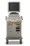
'Clinicians are demanding smaller, higher performing systems specifically designed to meet their clinical and operational challenges. The new Philips HD11 system provides an uncompromising platform, plus advanced options in a highly mobile and easy-to-use system.'
Device Information and Specification
APPLICATIONS
Abdominal, cardiac (also for adults with TEE), musculoskeletal (also pediatric), OB/GYN, prostate, smallparts, transcranial, vascular
CONFIGURATION
17' high resolution non-interlaced flat CRT, 4 active probe ports
B-mode, M-mode, coded harmonic imaging, color flow mode (CFM), power Doppler imaging (PDI), color Doppler, pulsed wave Doppler, tissue harmonic imaging
IMAGING OPTIONS
CrossXBeam spatial compounding, coded ultrasound acquisition),speckle reduction imaging (SRI), TruScan technology store raw data, CINE review with 4 speed types
OPTIONAL PACKAGE
Transesophageal scanning, stress echo, tissue velocity imaging (TVI), tissue velocity Doppler (TVD), contrast harmonic imaging
STORAGE, CONNECTIVITY, OS
Patient and image archive, HDD, DICOM 3.0, CD/DVD, MOD, Windows-based
DATA PROCESSING
Digital beamformer with 1024 system processing channel technology
H*W*D m (inch.)
1.62 * 0.61 * 0.99 (64 * 24 * 39)
WEIGHT
246 kg (498 lbs.)
POWER CONSUMPTION
less than 1.5 KVA
Result Pages : | Share This Page Look Ups |
Medical-Ultrasound-Imaging.com
former US-TIP.com
Member of SoftWays' Medical Imaging Group - MR-TIP • Radiology TIP • Medical-Ultrasound-Imaging
Copyright © 2008 - 2024 SoftWays. All rights reserved.
Terms of Use | Privacy Policy | Advertise With Us
former US-TIP.com
Member of SoftWays' Medical Imaging Group - MR-TIP • Radiology TIP • Medical-Ultrasound-Imaging
Copyright © 2008 - 2024 SoftWays. All rights reserved.
Terms of Use | Privacy Policy | Advertise With Us
[last update: 2023-11-06 01:42:00]