Medical Ultrasound Imaging
Wednesday, 8 May 2024
Info Sheets Out- side     | 'Directional Color Power Doppler' Searchterm 'Directional Color Power Doppler' found in 7 articles 1 term [ • ] - 3 definitions [• ] - 3 booleans [• ]Result Pages : • Directional Color Power Doppler
(DCPD) Directional color power Doppler combines power (amplitude) of Doppler signal with directional (phase) information to encode direction and variations in blood flow. DCPD is used in analysis of blood flow to assess blood vessel or vascular function. See also Color Power Doppler, Quadrature Detection, and Directional Indicators. •
(CPD) CPD is a type of color Doppler to visualize the presence of detectable blood flow. The flow information is based on the amplitude or strength of echoes received from moving cells and not on frequency shifts. Power Doppler is very sensitive to flowing blood but does not provide velocity or directional information. CPD is less angle dependent than traditional color Doppler, but more sensitive to motion artifacts. Color power angio (CPA) provides better sensitivity to slow flow states. The color maps for CPD are represented by a single continuous color (colour, Brit.). Because CPD does not provide directional information, no aliasing artifact occurs. See also Directional Color Power Doppler. Further Reading: Basics:
•
Color assignments can be switched on any ultrasound system and are used as directional indicators. The colors of red and blue indicates flow direction in color Doppler. Red is encoded to represent flows toward the transducer. Blue pixel encoding is used to depict flows away from the transducer. See also Directional Color Power Doppler. •
Doppler ultrasound is a medical imaging technique for calculating the relative velocity between two points by measuring the frequency shift of a sound wave transmitted from one point to the other, based on the Doppler effect. Continuous or pulsed Doppler is frequently used to examine cardiovascular blood flow. The combination of routine 2D-mode and Doppler ultrasound allows a complete evaluation of the heart's anatomy and function (including the fetal heart). See also Doppler Fluximetry in Pregnancy. Doppler ultrasound depends on the fact that if a moving object reflects the ultrasound waves, the echo frequencies are changed. A higher frequency is created if the object is moving toward the probe//transducer and a lower frequency if it is moving away from it. How much the frequency is changed depends upon how fast the object is moving. Doppler ultrasound shows the different rates of blood flow in different colors on a monitor in real time. The major Doppler parameters are the peak systolic velocity and the end-diastolic velocity. The peak systolic velocity ratio compensates the variability between different patients and instrumentations. Different Doppler and duplex techniques: Further Reading: News & More:
• 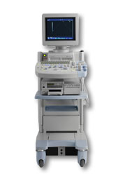 From Hitachi Medical Corporation (HMC), sales, marketing and service in the US by Hitachi Medical Systems America Inc.
The HI VISION™ 5500 - EUB-5500 fully digital ultrasound system delivers the latest generation of signal processing technology, sophisticated transducer design, and a host of features and options for advanced imaging capabilities across a wide range of clinical situations. This system is compatible with all Pentax ultrasound endoscopes.
Device Information and Specification
APPLICATIONS
Abdominal, brachytherapy/cryotherapy, breast, cardiac, dedicated biopsy, endoscopic, intraoperative, laparoscopic, musculoskeletal, OB/GYN, pediatric, small parts, urologic, vascular
CONFIGURATION
Compact system
Five frequency (except mini-probes)
Linear, convex, radial, miniradial/miniprobe, biplane, phased array, echoendoscope longitudinal, echoendoscope radial
3 modes of dynamic tissue harmonic imaging (dTHI), pulsed wave Doppler, continuous wave Doppler, color flow imaging, power Doppler, directional power Doppler, color flow angiography, real-time Doppler measurements
IMAGING OPTIONS
3RD generation color artifact suppression
OPTIONAL PACKAGE
STORAGE, CONNECTIVITY, OS
Patient and image database management system, HDD, FDD, MOD, CD-ROM, Network, DICOM 3.0, Windows XP
DATA PROCESSING
H*W*D m (inch.)
1.40 x 0.51 x 0.79 (55 x 20 x 31)
WEIGHT
130 kg (286 lbs.)
POWER CONSUMPTION
1.2kVA
ENVIRONMENTAL IMPACT
4096 btu/hr heat output
Result Pages : | Share This Page Look Ups |
Medical-Ultrasound-Imaging.com
former US-TIP.com
Member of SoftWays' Medical Imaging Group - MR-TIP • Radiology TIP • Medical-Ultrasound-Imaging
Copyright © 2008 - 2024 SoftWays. All rights reserved.
Terms of Use | Privacy Policy | Advertise With Us
former US-TIP.com
Member of SoftWays' Medical Imaging Group - MR-TIP • Radiology TIP • Medical-Ultrasound-Imaging
Copyright © 2008 - 2024 SoftWays. All rights reserved.
Terms of Use | Privacy Policy | Advertise With Us
[last update: 2023-11-06 01:42:00]