Medical Ultrasound Imaging
Wednesday, 8 May 2024
Info Sheets Out- side     | 'Brachytherapy' Searchterm 'Brachytherapy' found in 11 articles 1 term [ • ] - 10 definitions [• ] Result Pages : • Brachytherapy
Brachytherapy is a radiation therapy in which radioactive material (radioisotopes) sealed in needles, seeds or wires is placed directly into or near a tumor. Brachytherapy uses ultrasound imaging to visualize the needles for accurate placement of the small seeds or pellets (capsules) directly into e.g., the prostate. Ultrasound imaging allows accurate planning, placement and implantation of the radiation sources. Implantation of the seeds is a minimally invasive procedure. Radioactive seeds are inserted through the perineum skin (the area between the scrotum and the anus) into the prostate gland. With correct planning, the surgeon can implant the radiation sources for maximum benefits to effective cancer treatment. See also EchoSeed™, Prostate Ultrasound, Thermotherapy, High Intensity Focused Ultrasound, Urologic Ultrasound, Transurethral Sonography. • View NEWS results for 'Brachytherapy' (1). Further Reading: News & More:
•
Urologic ultrasound includes the examination of the kidneys, renal vessels, urinary tract, bladder, prostate, and scrotum. Usual gray scale ultrasound equipment and standard probes are sufficient to examine the kidney parenchyma and renal pelvis, the urinary tract and bladder. Doppler ultrasound is a useful adjunct to kidney ultrasound. High ultrasound system performance is desirable to show the arterial system, because advanced power Doppler is significantly more sensitive to blood flow than standard color Doppler. Transurethral sonography may be used to examine the bladder and urethra. Transrectal sonography is used to scan and treat the prostate e.g., with brachytherapy or high intensity focused ultrasound. Very small probes are used for these applications. Reflux sonography is especially used in pediatric ultrasound. See also Ultrasound Imaging Procedures, Ultrasound Picture, Ultrasound Imaging Modes, Lithotripsy, Thermotherapy, Brachytherapy and Ultrasound Therapy. Further Reading: Basics:
News & More:
• 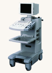 From Hitachi Medical Systems America Inc.;
The EUB-2000 B/W is a compact system capable of B-mode, M-mode, and optional with grayscale Doppler (EUB-2000 B/W Dop). This unit features intuitive operation, high mobility, and exceptional image quality. When color Doppler is not required, this system is an excellent choice for general purpose scanning. Device Information and Specification
APPLICATIONS
General purpose, OB/GYN, EUS, brachytherapy
CONFIGURATION
Compact system
Triple frequency
Linear, convex, radial, bi-plane, echoendoscope longitudinal
PROBE PORTS
Two
IMAGING OPTIONS
Simultaneous imaging with biplane probes
ADDITIONAL FEATURES
Cine / multi memory, application specific measuring, etc.
•
EchoSeed™ consists of radioactive 125I (iodine-125) seeds/implants indicated for the treatment of prostate cancer with brachytherapy. The design of this second generation product, used in combination with ultrasound imaging, facilitates optimal placement. EchoSeed™ allows the physician to view both the prostate gland and the seeds during the course of the implant procedure. Nycomed Amersham. Source: PR Newswire - 06/04/01. • 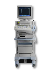 From Hitachi Medical Corporation (HMC), sales, marketing and service in the US by Hitachi Medical Systems America Inc.
The HI VISION™ 5500 - EUB-5500 fully digital ultrasound system delivers the latest generation of signal processing technology, sophisticated transducer design, and a host of features and options for advanced imaging capabilities across a wide range of clinical situations. This system is compatible with all Pentax ultrasound endoscopes.
Device Information and Specification
APPLICATIONS
Abdominal, brachytherapy/cryotherapy, breast, cardiac, dedicated biopsy, endoscopic, intraoperative, laparoscopic, musculoskeletal, OB/GYN, pediatric, small parts, urologic, vascular
CONFIGURATION
Compact system
Five frequency (except mini-probes)
Linear, convex, radial, miniradial/miniprobe, biplane, phased array, echoendoscope longitudinal, echoendoscope radial
3 modes of dynamic tissue harmonic imaging (dTHI), pulsed wave Doppler, continuous wave Doppler, color flow imaging, power Doppler, directional power Doppler, color flow angiography, real-time Doppler measurements
IMAGING OPTIONS
3RD generation color artifact suppression
OPTIONAL PACKAGE
STORAGE, CONNECTIVITY, OS
Patient and image database management system, HDD, FDD, MOD, CD-ROM, Network, DICOM 3.0, Windows XP
DATA PROCESSING
H*W*D m (inch.)
1.40 x 0.51 x 0.79 (55 x 20 x 31)
WEIGHT
130 kg (286 lbs.)
POWER CONSUMPTION
1.2kVA
ENVIRONMENTAL IMPACT
4096 btu/hr heat output
Result Pages : | Share This Page Look Ups |
Medical-Ultrasound-Imaging.com
former US-TIP.com
Member of SoftWays' Medical Imaging Group - MR-TIP • Radiology TIP • Medical-Ultrasound-Imaging
Copyright © 2008 - 2024 SoftWays. All rights reserved.
Terms of Use | Privacy Policy | Advertise With Us
former US-TIP.com
Member of SoftWays' Medical Imaging Group - MR-TIP • Radiology TIP • Medical-Ultrasound-Imaging
Copyright © 2008 - 2024 SoftWays. All rights reserved.
Terms of Use | Privacy Policy | Advertise With Us
[last update: 2023-11-06 01:42:00]