Medical Ultrasound Imaging
Sunday, 28 April 2024
Info Sheets Out- side     | 'Vascular Ultrasound' Searchterm 'Vascular Ultrasound' found in 29 articles 3 terms [ • ] - 10 definitions [• ] - 16 booleans [• ]Result Pages : • Vascular Ultrasound
Vascular ultrasound obtains images and measures blood flow velocity in the carotids, abdominal aorta, and vessels of kidneys, arms, or legs. Blockages in arteries, blood clots in veins, or abdominal aortic aneurysm can be detected. These abnormalities in blood flow are usually examined with different Doppler techniques. In addition, the speed and direction of blood flow can be color coded in a color map. Duplex techniques show both, the vessels and the surrounding tissue. The use of ultrasound contrast agents improves the left ventricular opacification in cardiac ultrasound examination. Usually, for a vascular ultrasound no special preparation is needed. See also Echocardiography, Venous Ultrasound, Adventitia, Intima, Temporal Mean Velocity, and Intravascular Ultrasound. Further Reading: News & More:
•
(IVUS) For intravascular ultrasound a small IVUS catheter with a probe is introduced into the artery. The transducer transmits and receives acoustic energy through this catheter. The reflected acoustic energy is used to build a picture of the inside of the vessel. A IVUS image consists of three layers around the lumen, the intima, media and adventitia. In addition, elastography or palpography could be used to evaluate the local mechanical properties of tissues (e.g. lipid pools in high-risk vulnerable atherosclerotic plaques). These techniques use the deformation caused by the intraluminal pressure generated by the probe. A high strain region at the lumen vessel wall boundary has 88% sensitivity and 89% specificity for identifying vulnerable plaques. There are high strain values of 1% in soft plaques with increased strain up to 2% at the shoulders of the plaque, while calcified material shows low strain values (0-0.2%). The radial strain in the tissue is obtained by cross-correlation techniques on the radio frequency signal. The strain is color-coded and plotted as a complimentary image to the intravascular ultrasound echogram. See also Interventional Ultrasound, Vascular Ultrasound. •
Vascular ultrasound contrast agents are gas microbubbles with a diameter less than 10 μm (2 to 5 μm on average for most of the newer agents) to pass through the lung capillaries and enter into the systemic circulation. Air bubbles in that size persist in solution for only a short time; too short for systemic vascular use in medical ultrasound imaging. So the gas bubbles have to be stabilized to persist long enough and survive pressure changes in the heart. Most vascular contrast media are stabilized against dissolution and coalescence by the presence of additional materials at the gas-liquid interface. In some cases, this material is an elastic solid shell that enhances stability by supporting a strain to counter the effect of surface tension. In other cases, the material is a surfactant, or a combination of two or more surfactants. Typically the effective duration of vascular enhancement is a few minutes, after which the microbubbles dissipate. This rather short duration of vascular enhancement makes it easy to perform repeated dynamic studies. Intravenous vascular contrast agents will be used in imaging malignant tumors in the liver, kidney, ovary, pancreas, prostate, and breast. Tumor neovascularization can be a marker for angiogenesis, and Doppler signals from small tumor vessels may be detectable after contrast injection. Contrast agents are useful for evaluating vessels in a variety of organs, including those involved in renal, hepatic, and pancreatic transplants. If an area of ischemia or a stenosis is detected after contrast administration, the use of other more expensive imaging modalities, including CT and MRI, can often be avoided. See also Acoustically Active Lipospheres. •  From Siemens Medical Systems;
We see a way to combine quality, versatility, and affordability in a slim, 20-inch wide, ergonomically designed ultrasound system. The new ACUSON CV70™ cardiovascular ultrasound system provides dedicated and complete cardiovascular capabilities in a powerful, all-digital ultrasound system offering exceptional image quality, Doppler sensitivity, color flow sensitivity and spatial resolution − everything you associate with the ACUSON family of cardiovascular products. Specifications for this system will be available soon. • 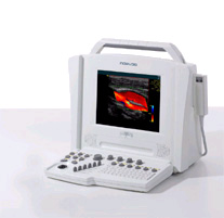 From Siemens Medical Systems;
We see a way to review 100% of your cardiovascular ultrasound exams and reports anywhere, anytime. The Cypress™ system is a highly miniaturized, all digital, phased array echocardiography system that provides complete studies and outstanding images - even on the most technically difficult patients. Specifications for this system will be available soon. Result Pages : | Share This Page Look Ups |
Medical-Ultrasound-Imaging.com
former US-TIP.com
Member of SoftWays' Medical Imaging Group - MR-TIP • Radiology TIP • Medical-Ultrasound-Imaging
Copyright © 2008 - 2024 SoftWays. All rights reserved.
Terms of Use | Privacy Policy | Advertise With Us
former US-TIP.com
Member of SoftWays' Medical Imaging Group - MR-TIP • Radiology TIP • Medical-Ultrasound-Imaging
Copyright © 2008 - 2024 SoftWays. All rights reserved.
Terms of Use | Privacy Policy | Advertise With Us
[last update: 2023-11-06 01:42:00]