Medical Ultrasound Imaging
Wednesday, 8 May 2024
Info Sheets Out- side     | 'Kidney Ultrasound' Searchterm 'Kidney Ultrasound' found in 15 articles 1 term [ • ] - 3 definitions [• ] - 11 booleans [• ]Result Pages : • Kidney Ultrasound
Ultrasonography of the kidneys (renal ultrasound) is part of a complete examination of the abdomen. Ultrasound is used to determine the size, shape, and exact position of the kidneys. Renal ultrasound provides important information regarding kidney function, related blood vessels, kidney stones, renal cysts, tumors, or hydronephrosis (suggestive of obstruction or blockage of the kidney). The kidneys are scanned on longitudinal and transverse planes. Patients should avoid carbonated drinks such as soda or seltzer the day before, and have a full bladder for the test. Lithotripsy is a therapeutic ultrasound procedure used to shatter simple stones in the kidney or upper urinary tract. See also Urologic Ultrasound, Reflux Sonography. •
(ESWL) Extracorporeal shock wave lithotripsy is a special use of kidney ultrasound, where high intensity focused ultrasound pulses are used to break up calcified stones in the kidney, bladder, or urethra. Pulses of sonic waves pulverize dense renal stones, which are then more easily passed through the ureter and out of the body in the urine. The ultrasound energy at high acoustic power levels is focused to a point exactly on the stone requiring an ultrasound scanning gel for maximum acoustic transmission. Air bubbles in the ultrasound couplant, regardless of their size, degrade the performance of Lithotripsy and have the following effect: Air bubbles smaller that 1/4 wavelength cause scattering of the sound waves as omni directional scatterers and less acoustic energy reaches the focal point. The result is less acoustic power at the focal point to disintegrate the kidney stone. Air bubbles larger than 1/4 wavelength act as reflectors and deflects the acoustic energy off in a different direction. These results in less acoustic energy at the focal point. Microbubbles dispersed throughout the ultrasound couplant layer change the average acoustic impedance of the gel layer (which reduces the total transmitted energy) and, due to refraction, change the focal point. Further Reading: News & More:
•
Reflux sonography, as an alternative to micturating cystography (MCU), evaluates vesico-ureteral reflux (VUR), a common problem in children. Contrast enhanced pulse-inversion imaging shows best results. During the instillation of an ultrasound contrast agent into the bladder, (as for a conventional MCU) the lower ureters and renal pelves are scanned transabdominally as the bladder is filled to stimulate micturition. Advantages for reflux sonography are a high sensitivity and the avoidance of X-rays. A disadvantage is the poorer depiction of the posterior urethra. However, for girls and for all follow-up studies, the ultrasound MCU has become standard in many pediatric ultrasound departments. See also Urologic Ultrasound, Kidney Ultrasound, Ultrasound Safety, Ultrasound Imaging Modes. •
Urologic ultrasound includes the examination of the kidneys, renal vessels, urinary tract, bladder, prostate, and scrotum. Usual gray scale ultrasound equipment and standard probes are sufficient to examine the kidney parenchyma and renal pelvis, the urinary tract and bladder. Doppler ultrasound is a useful adjunct to kidney ultrasound. High ultrasound system performance is desirable to show the arterial system, because advanced power Doppler is significantly more sensitive to blood flow than standard color Doppler. Transurethral sonography may be used to examine the bladder and urethra. Transrectal sonography is used to scan and treat the prostate e.g., with brachytherapy or high intensity focused ultrasound. Very small probes are used for these applications. Reflux sonography is especially used in pediatric ultrasound. See also Ultrasound Imaging Procedures, Ultrasound Picture, Ultrasound Imaging Modes, Lithotripsy, Thermotherapy, Brachytherapy and Ultrasound Therapy. Further Reading: Basics:
News & More:
• 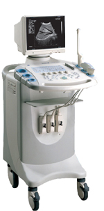
From SIUI Inc.;
Device Information and Specification
CONFIGURATION
Normal system, gray scale(256)
Linear and convex
PROBES STANDARD
2.5MHz ~ 11.0MHz, broad band, tri-frequency
IMAGING OPTIONS
Real zoom, max. Zoom * 4.0, multiplying power and position selectable
OPTIONAL PACKAGE
H*W*D m
1.30 * 0.52 * 0.77
WEIGHT
90kg (main unit)
POWER REQUIREMENT
AC 220V/110V, 50Hz/60Hz
POWER CONSUMPTION
0.45 KVA
Result Pages : | Share This Page Look Ups |
Medical-Ultrasound-Imaging.com
former US-TIP.com
Member of SoftWays' Medical Imaging Group - MR-TIP • Radiology TIP • Medical-Ultrasound-Imaging
Copyright © 2008 - 2024 SoftWays. All rights reserved.
Terms of Use | Privacy Policy | Advertise With Us
former US-TIP.com
Member of SoftWays' Medical Imaging Group - MR-TIP • Radiology TIP • Medical-Ultrasound-Imaging
Copyright © 2008 - 2024 SoftWays. All rights reserved.
Terms of Use | Privacy Policy | Advertise With Us
[last update: 2023-11-06 01:42:00]