Medical Ultrasound Imaging
Wednesday, 8 May 2024
Info Sheets Out- side     | 'M-Mode' Searchterm 'M-Mode' found in 47 articles 2 terms [ • ] - 45 definitions [• ] Result Pages : • M-Mode
The M-mode (Motion-mode) ultrasound is used for analyzing moving body parts (also called time-motion or TM-mode) commonly in cardiac and fetal cardiac imaging. The application of B-mode and a strip chart recorder allows visualization of the structures as a function of depth and time. The M-mode ultrasound transducer beam is stationary while the echoes from a moving reflector are received at varying times.
A single beam in an ultrasound scan is used to produce the one-dimensional M-mode picture, where movement of a structure such as a heart valve can be depicted in a wave-like manner. The high sampling frequency (up to 1000 pulses per second) is useful in assessing rates and motion, particularly in cardiac structures such as the various valves and the chamber walls. Further Reading: News & More:
•
M-mode (Motion-mode) ultrasound shows the motion of cardiac structures. M-mode echocardiography records the amplitude and rate of motion of a moving structure in real time by repeatedly measuring the distance of the object from the single transducer at a given moment. The single sound beam is transmitted and reflected signals are displayed as dots of varying intensities, creating lines across the screen. It yields a one-dimensional image, sometimes called an 'ice pick' view of the heart. M-mode echocardiography is used to detect valvulopathies (calcifications, etc.) and cardiomyopathies (dyskinesis, aneurysm, etc.). See also Bicycle Stress Echocardiography, Transthoracic Echocardiography, and Transesophageal Echocardiography. Further Reading: News & More: •
Cardiac ultrasound, also known as echocardiography or echocardiogram, is used to provide several different levels and types of heart testing. Cardiac ultrasound utilizes the same ultrasound principles as used for obstetric and gynecologic evaluations of pregnant women, gallbladder ultrasound and other abdominal structures. The ultrasound is directed out of a hand held probe which can be moved to image the heart from different positions. Additionally, so that heart events can be timed, ECG leads are placed on the chest. The reflected wave is converted into an actual image of the heart and displayed in a real-time mode or M-mode ultrasound format. M-mode recordings permit measurement of cardiac dimensions and detailed analysis of complex motion patterns depending on transducer angulations. Also the time relationships with other physiological variables such as ECG, heart sounds, and pulse tracings, can be recorded simultaneously. A stress echocardiogram provides information about the cardiac performance. Two-dimensional tomographic images of selected cardiac sections give more information than M-mode about the shape of the heart and also show the spatial relationships of its structures during the cardiac cycle (diastole to systole). See also M-Mode Echocardiography, and Myocardial Contrast Echocardiography. Further Reading: News & More:
•  From SIUI Inc.;
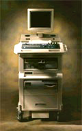
'The Apogee 800Plus ultrasound system offers a new level of high quality in image. With broad clinical application, the system delivers ease of use for cardiac and vascular exams.' 'Due to the exceptional image quality, the sensitivity of color Doppler and many other advanced features, the Apogee 800Plus offers complete diagnosis capability for your entire patient population, including abdominal, obstetrics, gynecology, adult cardiology, pediatric cardiology, peripheral vascular, small parts, breast, prostate, transcranial, etc.'
Device Information and Specification
APPLICATIONS
Linear, curved, phased and symmetric phased array
2D, M-mode, Pulsed wave Doppler, Color Power Angio Imaging, Color 2D, Color M-mode, High PRF Doppler
H*W*D m
1.46 * 0.69 * 0.87
WEIGHT
140 kg (without peripherals)
POWER REQUIREMENT
115Vac, (V~), @ 50 to 60 Hz, 1.2kVA
230Vac, (V~), @ 50 to 60 Hz, 1.2kVA POWER CONSUMPTION
800 Watts (with optional OEM'S: 1200 Watts max)
• 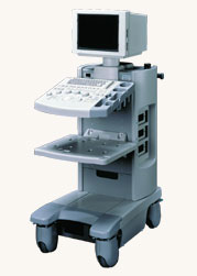 From Hitachi Medical Systems America Inc.;
The EUB-2000 B/W is a compact system capable of B-mode, M-mode, and optional with grayscale Doppler (EUB-2000 B/W Dop). This unit features intuitive operation, high mobility, and exceptional image quality. When color Doppler is not required, this system is an excellent choice for general purpose scanning. Device Information and Specification
APPLICATIONS
General purpose, OB/GYN, EUS, brachytherapy
CONFIGURATION
Compact system
Triple frequency
Linear, convex, radial, bi-plane, echoendoscope longitudinal
PROBE PORTS
Two
IMAGING OPTIONS
Simultaneous imaging with biplane probes
ADDITIONAL FEATURES
Cine / multi memory, application specific measuring, etc.
Result Pages : | Share This Page Look Ups |
Medical-Ultrasound-Imaging.com
former US-TIP.com
Member of SoftWays' Medical Imaging Group - MR-TIP • Radiology TIP • Medical-Ultrasound-Imaging
Copyright © 2008 - 2024 SoftWays. All rights reserved.
Terms of Use | Privacy Policy | Advertise With Us
former US-TIP.com
Member of SoftWays' Medical Imaging Group - MR-TIP • Radiology TIP • Medical-Ultrasound-Imaging
Copyright © 2008 - 2024 SoftWays. All rights reserved.
Terms of Use | Privacy Policy | Advertise With Us
[last update: 2023-11-06 01:42:00]