Medical Ultrasound Imaging
Wednesday, 8 May 2024
Info Sheets Out- side     | 'Continuous Wave Doppler' Searchterm 'Continuous Wave Doppler' found in 18 articles 1 term [ • ] - 14 definitions [• ] - 3 booleans [• ]Result Pages : • Continuous Wave Doppler
(CWD) Continuous wave (CW) Doppler is an ultrasound imaging mode, which records blood flow velocities along the length of the beam. Continuous wave Doppler uses different crystals to send and receive the signal. The transducer operating in continuous wave mode utilizes one half of the elements and is continuously sending sound waves of a single frequency while the other half is continuously receiving the reflected signals. The advantages of a continuous wave transducer are a high sensitivity and no Nyquist limit. CW Doppler does not alias but has no depth precision and large gate. The beat frequency is the Doppler shift. CW Doppler echocardiography employs this technique to record the flow of blood through the cardiovascular system. See also Cross Talk, Periorbital Doppler, and Mirror Artifact. Further Reading: News & More:
•
Cross talk is an ultrasound artifact in which strong sound signals in one directional channel leak into another, appearing as a mirror image of the spectral display on the opposite side of the baseline. Cross talk distinguishes the condition of undesired crossover of transmitted sound waves into the receiving transducer in a continuous wave Doppler system. Also called Mirror Artifact. •
Doppler techniques are dependent on the transducers used. The transducer operating in continuous wave mode utilizes one half of the elements and is continuously sending sound energy while the other half is continuously receiving the reflected signals. If the transducer is being used in a pulsed wave mode, the whole transducer is used to send and then receive the returning signals. Pulsed wave techniques allow the accurate measurement of blood flow at a specific area in the heart and the detection of both velocity and direction. Measurement is performed by timing the reception of the returning signals giving a view of flows at specific depths. The region where flow velocities are measured is called the sample volume. Errors in the accuracy of the information arise if the velocities exceed a certain speed. The highest velocity accurately measured is called the Nyquist limit.
•
Continuous Wave Doppler
Used for accurate measurement of high Velocity flow. A disadvantage is the poor range of resolution.
•
Pulsed Wave Doppler
Used for the measurement of velocities at a specific location with a good range of resolution. A disadvantage is the imprecise measuring of high velocities. See also Doppler Velocity Signal and Doppler Effect. Further Reading: Basics:
News & More:
•
Doppler ultrasound is a medical imaging technique for calculating the relative velocity between two points by measuring the frequency shift of a sound wave transmitted from one point to the other, based on the Doppler effect. Continuous or pulsed Doppler is frequently used to examine cardiovascular blood flow. The combination of routine 2D-mode and Doppler ultrasound allows a complete evaluation of the heart's anatomy and function (including the fetal heart). See also Doppler Fluximetry in Pregnancy. Doppler ultrasound depends on the fact that if a moving object reflects the ultrasound waves, the echo frequencies are changed. A higher frequency is created if the object is moving toward the probe//transducer and a lower frequency if it is moving away from it. How much the frequency is changed depends upon how fast the object is moving. Doppler ultrasound shows the different rates of blood flow in different colors on a monitor in real time. The major Doppler parameters are the peak systolic velocity and the end-diastolic velocity. The peak systolic velocity ratio compensates the variability between different patients and instrumentations. Different Doppler and duplex techniques: Further Reading: News & More:
• 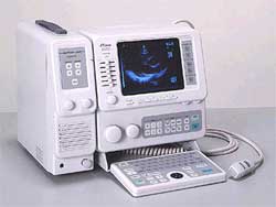 From Fukuda Denshi Co., Ltd.;
'Phased array probes made available for High resolution imaging of full cardiovascular examinations. By adding Linear and Convex probes, the examination areas are extended to Abdominal and OB/GYN applications. B-mode (1, 2 displays), M, and B/M-modes can be individually shown. Optional Doppler unit available for observation of Blood flow information in pulsed and continuous wave Doppler. Portable with IP (Image Processing), Full Keyboard, and track ball are standard.' Result Pages : | Share This Page Look Ups |
Medical-Ultrasound-Imaging.com
former US-TIP.com
Member of SoftWays' Medical Imaging Group - MR-TIP • Radiology TIP • Medical-Ultrasound-Imaging
Copyright © 2008 - 2024 SoftWays. All rights reserved.
Terms of Use | Privacy Policy | Advertise With Us
former US-TIP.com
Member of SoftWays' Medical Imaging Group - MR-TIP • Radiology TIP • Medical-Ultrasound-Imaging
Copyright © 2008 - 2024 SoftWays. All rights reserved.
Terms of Use | Privacy Policy | Advertise With Us
[last update: 2023-11-06 01:42:00]