Medical Ultrasound Imaging
Wednesday, 8 May 2024
Info Sheets Out- side     | 'Tissue Harmonic Imaging' Searchterm 'Tissue Harmonic Imaging' found in 21 articles 1 term [ • ] - 20 definitions [• ] Result Pages : • Tissue Harmonic Imaging
(THI) Tissue harmonic imaging (also called native harmonic imaging) is a signal processing technique which addresses ultrasound limitations like penetration and resolution. Tissue harmonic imaging reduces noise and clutter by improving signal to noise ratio and resolution. The signal penetration in soft tissue increases as the transmit frequency is decreased, by simultaneous decreased image resolution. As an ultrasound wave propagates through the target media a change occurs in the shape and frequency of the transmitted signal. The change is due to the normal resistance of tissue to propagate sound energy. This resistance and the resulting signal change is called a harmonic oscillation. For harmonic imaging the input frequency doubles the output frequency, for example a transmit frequency of 3.0 MHz. which would provide maximum penetration will return a harmonic frequency of 6.0 MHz. The returning higher frequency signal has to only travel one direction to the probe. The advantages of high frequency imaging and the one-way travel effect are decreased reverberation, beam aberration, and side lobes, as well as increased resolution and cystic clearing. • 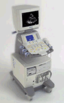 From Siemens Medical Systems;
'This high-performance, multi-specialty system supports and improves your daily ultrasound routine. Embedded DICOM creates the integrated foundation for a complete connectivity solution while MultiHertz™ multiple frequency imaging and Tissue Harmonic Imaging (THI) expands the clinical versatility of the system. Advanced applications such as stress echo, SieScape™ panoramic imaging, and transesophageal imaging can be seamlessly integrated.'
Device Information and Specification
CLINICAL APPLICATION
Widest range of applications
CONFIGURATION
Compact, mobile system
Wideband MultiHertz™ multiple frequency
IMAGING OPTIONS
OPTIONAL PACKAGE
Software upgradeability to advanced clinical application
IMAGING ENHANCEMENTS
Precision MotionCapture, Synthetic aperture technology
STORAGE
Patient and image database management system
DATA PROCESSING
Parallel and quad signal processing
WEIGHT
Lightweight
•  From Siemens Medical Systems;
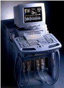
The new gold standard in echocardiography is defined by Native Tissue Harmonic Imaging (NTHI) and DELTA Differential Echo Amplification−two powerful technologies that were first introduced on Acuson's Sequoia™ echocardiography system. Now they have migrated to the Aspen™ Advanced system. Specifications for this system will be available soon. • 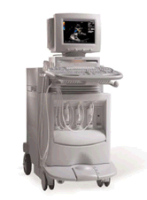 From Siemens Medical Systems;
'Conventional ultrasound systems are unable to compensate for the unique acoustic signature of individual patients. But there is nothing conventional about the new ACUSON Sequoia™ C512 echocardiography system with Sequoia™ matched response technology.'
Device Information and Specification
APPLICATIONS
CONFIGURATION
Compact, portable
2D-Mode, M-mode, Cadence™, Native® Tissue Harmonic Imaging, Transmit Compounding, SST™ Color and Solo™ Spectral Doppler
STORAGE, CONNECTIVITY, OS
KinetDx integrated PACS, DIMAQ-Workstation
•  From ALOKA Co., Ltd.;
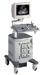
'A Platform for Digital, Pure-Beam Imaging The high-performance, ALOKA ProSound SSD-3500 utilizes advanced ProSound technologies including: Fully digital beam former A wide dynamic range, 12-bit A/D converter Multi beam processing. The SSD-3500 also helps you achieve more efficient examinations. Its ergonomic, user-friendly design enables you to customize the system according to your specific application needs.'
Device Information and Specification
APPLICATIONS
CONFIGURATION
Compact, portable, dual dynamic display
Color Flow, Power Flow, Spectral Doppler, Real-time Free Angular M-Mode, Tissue Harmonic Imaging, Quint Frequency Imaging, Pure Harmonic Detection
STORAGE, CONNECTIVITY, OS
Data Management Subsystem (iDMS), DICOM-Worklist
DATA PROCESSING
12-bit analog to digital converter
Result Pages : | Share This Page Look Ups |
Medical-Ultrasound-Imaging.com
former US-TIP.com
Member of SoftWays' Medical Imaging Group - MR-TIP • Radiology TIP • Medical-Ultrasound-Imaging
Copyright © 2008 - 2024 SoftWays. All rights reserved.
Terms of Use | Privacy Policy | Advertise With Us
former US-TIP.com
Member of SoftWays' Medical Imaging Group - MR-TIP • Radiology TIP • Medical-Ultrasound-Imaging
Copyright © 2008 - 2024 SoftWays. All rights reserved.
Terms of Use | Privacy Policy | Advertise With Us
[last update: 2023-11-06 01:42:00]