Medical Ultrasound Imaging
Wednesday, 8 May 2024
Info Sheets Out- side     | 'Contrast Harmonic Imaging' Searchterm 'Contrast Harmonic Imaging' found in 24 articles 1 term [ • ] - 8 definitions [• ] - 15 booleans [• ]Result Pages : • Contrast Harmonic Imaging
(CHI) Contrast harmonic imaging is an ultrasound technique to improve the measurement of blood perfusion or capillary blood flow. Based on the nonlinear properties of contrast agents, CHI transmits at the fundamental frequency but receives at the second harmonic. Contrast enhanced echo signals contain significant energy components at higher harmonics (bubbles acts as harmonic oscillators), while tissue echoes do not. Caused by that contrast signal can be separated from tissue echoes by the characteristic signal. In combination with the pulse inversion technique, CHI promises very high contrast agent sensitivity with high spatial resolution. See also Ultrasound Contrast Agent Safety and Hemoglobin. Further Reading: Basics:
•
(CEUS) Contrast agents increase the reflection of ultrasonic energy, improve the signal to noise ratio and caused by that the detection of abnormal microvascular and macrovascular disorders. Contrast enhanced ultrasound is used in abdominal ultrasound (liver sonography) as well as in cerebrovascular examinations e.g., for an accurate grading of carotid stenosis. The used contrast agents are safe and well tolerated. The quality of the enhancement depends on:
•
the concentration of the contrast agent;
•
the type of injection, flow rate;
•
the patient characteristics;
•
the microbubble quality and properties of the filling gas and the shell.
The additional use of ultrasound contrast agents (USCAs) may overcome typical limitations like poor contrast of B-mode imaging or limited sensitivity of Doppler techniques. The development of new ultrasound applications (e.g., blood flow imaging, perfusion quantification) depends also from the development of pulse sequences for bubble specific imaging. In addition, contrast enhanced ultrasound improves the monitoring of ultrasound guided interventions like RF thermal ablation. See also Contrast Enhanced Doppler Imaging, Contrast Harmonic Imaging, Contrast Imaging Techniques and Contrast Pulse Sequencing. Further Reading: News & More:
•
Many different contrast imaging techniques have been developed. Most are either variations, hybrids, or combinations of the following ultrasound techniques: See also Coherent Contrast Imaging, Ultrasound Picture and Targeted Contrast Imaging. Further Reading: Basics:
•  From Philips Medical Systems;
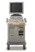
'Clinicians are demanding smaller, higher performing systems specifically designed to meet their clinical and operational challenges. The new Philips HD11 system provides an uncompromising platform, plus advanced options in a highly mobile and easy-to-use system.'
Device Information and Specification
APPLICATIONS
Abdominal, cardiac (also for adults with TEE), musculoskeletal (also pediatric), OB/GYN, prostate, smallparts, transcranial, vascular
CONFIGURATION
17' high resolution non-interlaced flat CRT, 4 active probe ports
B-mode, M-mode, coded harmonic imaging, color flow mode (CFM), power Doppler imaging (PDI), color Doppler, pulsed wave Doppler, tissue harmonic imaging
IMAGING OPTIONS
CrossXBeam spatial compounding, coded ultrasound acquisition),speckle reduction imaging (SRI), TruScan technology store raw data, CINE review with 4 speed types
OPTIONAL PACKAGE
Transesophageal scanning, stress echo, tissue velocity imaging (TVI), tissue velocity Doppler (TVD), contrast harmonic imaging
STORAGE, CONNECTIVITY, OS
Patient and image archive, HDD, DICOM 3.0, CD/DVD, MOD, Windows-based
DATA PROCESSING
Digital beamformer with 1024 system processing channel technology
H*W*D m (inch.)
1.62 * 0.61 * 0.99 (64 * 24 * 39)
WEIGHT
246 kg (498 lbs.)
POWER CONSUMPTION
less than 1.5 KVA
•
The earliest introduction of vascular ultrasound contrast agents (USCA) was by Gramiak and Shah in 1968, when they injected agitated saline into the ascending aorta and cardiac chambers during echocardiographic to opacify the left heart chamber. Strong echoes were produced within the heart, due to the acoustic mismatch between free air microbubbles in the saline and the surrounding blood.
•
In 1880 the Curie brothers discovered the piezoelectric effect in quartz. Converse piezoelectricity was mathematically deduced from fundamental thermodynamic principles by Lippmann in 1881.
•
In 1917, Paul Langevin (France) and his coworkers developed an underwater sonar system (called hydrophone) that uses the piezoelectric effect to detect submarines through echo location.
•
In 1935, the first RADAR system was produced by the British physicist Robert Watson-Wat. Also about 1935, developments began with the objective to use ultrasonic power therapeutically, utilizing its heating and disruptive effects on living tissues. In 1936, Siemens markets the first ultrasonic therapeutic machine, the Sonostat.
•
Shortly after the World War II, researchers began to explore medical diagnostic capabilities of ultrasound. Karl Theo Dussik (Austria) attempted to locate the cerebral ventricles by measuring the transmission of ultrasound beam through the skull. Other researchers try to use ultrasound to detect gallstones, breast masses, and tumors. These first investigations were performed with A-mode.
•
Shortly after the World War II, researchers in Europe, the United States and Japan began to explore medical diagnostic capabilities of ultrasound. Karl Theo Dussik (Austria) attempted to locate the cerebral ventricles by measuring the transmission of ultrasound beam through the skull. Other researchers, e.g. George Ludwig (United States) tried to use ultrasound to detect gallstones, breast masses, and tumors. This first experimentally investigations were performed with A-mode. Ultrasound pioneers contributed innovations and important discoveries, for example the velocity of sound transmission in animal soft tissues with a mean value of 1540 m/sec (still in use today), and determined values of the optimal scanning frequency of the ultrasound transducer.
•
In the early 50`s the first B-mode images were obtained. Images were static, without gray-scale information in simple black and white and compound technique. Carl Hellmuth Hertz and Inge Edler (Sweden) made in 1953 the first scan of heart activity. Ian Donald and Colleagues (Scotland) were specialized on obstetric and gynecologic ultrasound research. By continuous development it was possible to study pregnancy and diagnose possible complications.
•
After about 1960 two-dimensional compound procedures were developed. The applications in obstetric and gynecologic ultrasound boomed worldwide from the mid 60's with both, A-scan and B-scan equipment. In the late 60's B-mode ultrasonography replaced A-mode in wide parts.
•
In the 70's gray scale imaging became available and with progress of computer technique ultrasonic imaging gets better and faster.
•
•
In the 90's, high resolution scanners with digital beamforming, high transducer frequencies, multi-channel focus and broad-band transducer technology became state of the art. Optimized tissue contrast and improved diagnostic accuracy lead to an important role in breast imaging and cancer detection. Color Doppler and Duplex became available and sensitivity for low flow was continuously improved.
•
Actually, machines with advanced ultrasound system performance are equipped with realtime compound imaging, tissue harmonic imaging, contrast harmonic imaging, vascular assessment, matrix array transducers, pulse inversion imaging, 3D and 4D ultrasound with panoramic view.
Result Pages : | Share This Page Look Ups |
Medical-Ultrasound-Imaging.com
former US-TIP.com
Member of SoftWays' Medical Imaging Group - MR-TIP • Radiology TIP • Medical-Ultrasound-Imaging
Copyright © 2008 - 2024 SoftWays. All rights reserved.
Terms of Use | Privacy Policy | Advertise With Us
former US-TIP.com
Member of SoftWays' Medical Imaging Group - MR-TIP • Radiology TIP • Medical-Ultrasound-Imaging
Copyright © 2008 - 2024 SoftWays. All rights reserved.
Terms of Use | Privacy Policy | Advertise With Us
[last update: 2023-11-06 01:42:00]