Medical Ultrasound Imaging
Saturday, 27 April 2024
Info Sheets Out- side     | 'A-Mode' Searchterm 'A-Mode' found in 10 articles 1 term [ • ] - 9 definitions [• ] Result Pages : • A-Mode
A-mode (Amplitude-mode) ultrasound is a technique used to assess organ dimensions and determine the depth of an organ. While A-mode technology was previously employed in midline echoencephalography for rapid screening of intracranial mass lesions and ophthalmologic scanning, it is now considered obsolete in medical imaging. Nonetheless, the A-mode scan has found applications in early pregnancy assessment (specifically the detection of fetal heartbeats), cephalometry, and placental localization.
When the ultrasound beam encounters an anatomic boundary, the received sound impulse is processed to appear as a vertical reflection of a point. On the display, it looks like spikes of different heights (the amplitude). The intensity of the returning impulse determined the height of the vertical reflection and the time it took for the impulse to make the round trip would determine the space between verticals. The distance between these spikes can be measured accurately by dividing the speed of sound in tissue (1540 m/sec) by half the sound travel time. During an echoencephalography scan, the first A-mode scan is acquired from the right side of the head and captured on film. Subsequently, the probe is positioned at the corresponding point on the left side, and a second exposure is captured on the same film, displaying inverted spikes. The A-mode ultrasound could be used to identify structures normally located in the midline of the brain such as the third ventricle and falx cerebri. The midline structures would be aligned in normal patients but show displacement in patients with mass lesion such as a subdural, epidural, or intracranial hemorrhage. See also 2D Ultrasound, 3D Ultrasound, 4D Ultrasound, Ultrasound Biomicroscopy, A-scan, B-mode and the Infosheet about ultrasound modes. •
The earliest introduction of vascular ultrasound contrast agents (USCA) was by Gramiak and Shah in 1968, when they injected agitated saline into the ascending aorta and cardiac chambers during echocardiographic to opacify the left heart chamber. Strong echoes were produced within the heart, due to the acoustic mismatch between free air microbubbles in the saline and the surrounding blood.
•
In 1880 the Curie brothers discovered the piezoelectric effect in quartz. Converse piezoelectricity was mathematically deduced from fundamental thermodynamic principles by Lippmann in 1881.
•
In 1917, Paul Langevin (France) and his coworkers developed an underwater sonar system (called hydrophone) that uses the piezoelectric effect to detect submarines through echo location.
•
In 1935, the first RADAR system was produced by the British physicist Robert Watson-Wat. Also about 1935, developments began with the objective to use ultrasonic power therapeutically, utilizing its heating and disruptive effects on living tissues. In 1936, Siemens markets the first ultrasonic therapeutic machine, the Sonostat.
•
Shortly after the World War II, researchers began to explore medical diagnostic capabilities of ultrasound. Karl Theo Dussik (Austria) attempted to locate the cerebral ventricles by measuring the transmission of ultrasound beam through the skull. Other researchers try to use ultrasound to detect gallstones, breast masses, and tumors. These first investigations were performed with A-mode.
•
Shortly after the World War II, researchers in Europe, the United States and Japan began to explore medical diagnostic capabilities of ultrasound. Karl Theo Dussik (Austria) attempted to locate the cerebral ventricles by measuring the transmission of ultrasound beam through the skull. Other researchers, e.g. George Ludwig (United States) tried to use ultrasound to detect gallstones, breast masses, and tumors. This first experimentally investigations were performed with A-mode. Ultrasound pioneers contributed innovations and important discoveries, for example the velocity of sound transmission in animal soft tissues with a mean value of 1540 m/sec (still in use today), and determined values of the optimal scanning frequency of the ultrasound transducer.
•
In the early 50`s the first B-mode images were obtained. Images were static, without gray-scale information in simple black and white and compound technique. Carl Hellmuth Hertz and Inge Edler (Sweden) made in 1953 the first scan of heart activity. Ian Donald and Colleagues (Scotland) were specialized on obstetric and gynecologic ultrasound research. By continuous development it was possible to study pregnancy and diagnose possible complications.
•
After about 1960 two-dimensional compound procedures were developed. The applications in obstetric and gynecologic ultrasound boomed worldwide from the mid 60's with both, A-scan and B-scan equipment. In the late 60's B-mode ultrasonography replaced A-mode in wide parts.
•
In the 70's gray scale imaging became available and with progress of computer technique ultrasonic imaging gets better and faster.
•
•
In the 90's, high resolution scanners with digital beamforming, high transducer frequencies, multi-channel focus and broad-band transducer technology became state of the art. Optimized tissue contrast and improved diagnostic accuracy lead to an important role in breast imaging and cancer detection. Color Doppler and Duplex became available and sensitivity for low flow was continuously improved.
•
Actually, machines with advanced ultrasound system performance are equipped with realtime compound imaging, tissue harmonic imaging, contrast harmonic imaging, vascular assessment, matrix array transducers, pulse inversion imaging, 3D and 4D ultrasound with panoramic view.
•
A-scans are used in ophthalmologic scanning, to detect and monitor pregnancy problems, and screen intracranial mass lesions by using A-modes. A-scan ultrasound biometry, commonly referred to as an A-scan, is a routine diagnostic test used in ophthalmology. The A-scan provides data on the shape of the eye, which is a major determinant in common sight disorders. Ultrasound scanners used in this type of test require usually direct contact with the eye. See also A-Mode, Oculoplethysmography, Ultrasound Biomicroscopy, B-Scan, C-Scan and D-Scan. Further Reading: News & More:
•
Also called B-mode echography, B-mode sonography, 2D-mode, and sonogram. B-mode ultrasound (Brightness-mode) is the display of a 2D-map of B-mode data, currently the most common form of ultrasound imaging. The development from A-mode to B-mode is that the ultrasound signal is used to produce various points whose brightness depends on the amplitude instead of the spiking vertical movements in the A-mode. Sweeping a narrow ultrasound beam through the area being examined while transmitting pulses and detecting echoes along closely spaced scan lines produces B-scan images. The vertical position of each bright dot is determined by the time delay from pulse transmission to return of the echo, and the horizontal position by the location of the receiving transducer element. To generate a rapid series of individual 2D images that show motion, the ultrasound beam is swept repeatedly. The returning sound pulses in B-mode have different shades of darkness depending on their intensities. The varying shades of gray reflect variations in the texture of internal organs. This form of display (solid areas appear white and fluid areas appear black) is also called gray scale. Different types of displayed B-mode images are: The probe movement can be performed manual (compound and static B-scanner) or automatic (real-time scanner). The image reconstruction can be parallel or sector type. See also B-Scan, 4B-Mode, and Harmonic B-Mode Imaging. Further Reading: News & More:
• 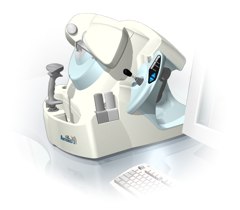 From Ultralink LLC;
'Artemis is a very high frequency (VHF) ultrasound eye scanner. In use, the patient leans forward placing their head onto an adjustable headrest. The headrest's unique design permits the patient to pull away quickly from the scanner if desired. An eyecup filled with a saline-based interface fluid couples the ultrasound signal to the eye, while a precision mechanism moves the transducer past the front of the eye. During the accurately controlled arc motion of the transducer, which lasts less than one second, many thousands of ultrasound samples are digitized. Following a scan, signal analysis is performed on a PC-compatible microcomputer, and the data are available for immediate viewing on an LCD monitor or disk storage. Artemis is very flexible; many adjustments to the scanning parameters are possible to customize the scan to your clinical needs. Functions are provided for centering the scan about the optical axis of the eye. The starting location of the scans as well as the extent can be varied as desired, to view image planes through the eye at different angles.' See also Ultrasound Biomicroscopy, A-Mode and A-Scan. Further Reading: News & More:
Result Pages : | Share This Page Look Ups |
Medical-Ultrasound-Imaging.com
former US-TIP.com
Member of SoftWays' Medical Imaging Group - MR-TIP • Radiology TIP • Medical-Ultrasound-Imaging
Copyright © 2008 - 2024 SoftWays. All rights reserved.
Terms of Use | Privacy Policy | Advertise With Us
former US-TIP.com
Member of SoftWays' Medical Imaging Group - MR-TIP • Radiology TIP • Medical-Ultrasound-Imaging
Copyright © 2008 - 2024 SoftWays. All rights reserved.
Terms of Use | Privacy Policy | Advertise With Us
[last update: 2023-11-06 01:42:00]