Medical Ultrasound Imaging
Thursday, 9 May 2024
'Compound B-Mode' Searchterm 'Compound B-Mode' found in 12 articles 1 term [ • ] - 3 definitions [• ] - 8 booleans [• ]Result Pages : • Compound B-Mode
Compound B-mode imaging takes different forms and refers to different methods of creating the ultrasound image. Real-time compound ultrasound improves the image quality of B-mode scanning by combining ultrasound information obtained from multiple angles. The used averaging process of compound B-mode reduces artifacts and improves the representation of true image data. B-mode images and Doppler mode images (see also Duplex) can be compounded on the display to improve the visualization of the anatomical relationships between vessels and the surrounding tissues. Further Reading: News & More:
•
Real-time mode has been developed to present motion like a movie of the body's inner workings, showing this information at a high rate. The special real-time transducer uses a larger sound beam than for A, B or M-modes. A linear array transducer with multiple crystal elements displays real-time compound B-mode images with up to 100 images per second. At each scan line, one sound pulse is transmitted and all echoes from the surface to the deepest range are received. Then the ultrasound beam moves on to the next scan line position where pulse transmission and echo recording are repeated. See also Compound B-Mode, Pulse Inversion Doppler, and Frame Averaging. Further Reading: News & More:
•
Also called B-mode echography, B-mode sonography, 2D-mode, and sonogram. B-mode ultrasound (Brightness-mode) is the display of a 2D-map of B-mode data, currently the most common form of ultrasound imaging. The development from A-mode to B-mode is that the ultrasound signal is used to produce various points whose brightness depends on the amplitude instead of the spiking vertical movements in the A-mode. Sweeping a narrow ultrasound beam through the area being examined while transmitting pulses and detecting echoes along closely spaced scan lines produces B-scan images. The vertical position of each bright dot is determined by the time delay from pulse transmission to return of the echo, and the horizontal position by the location of the receiving transducer element. To generate a rapid series of individual 2D images that show motion, the ultrasound beam is swept repeatedly. The returning sound pulses in B-mode have different shades of darkness depending on their intensities. The varying shades of gray reflect variations in the texture of internal organs. This form of display (solid areas appear white and fluid areas appear black) is also called gray scale. Different types of displayed B-mode images are: The probe movement can be performed manual (compound and static B-scanner) or automatic (real-time scanner). The image reconstruction can be parallel or sector type. See also B-Scan, 4B-Mode, and Harmonic B-Mode Imaging. Further Reading: News & More:
•
Duplex ultrasonography (duplex scan) consists of two ultrasound modalities to study blood flow and the perivascular tissue. This includes B-mode / gray scale imaging used in combination with spectral Doppler / pulsed-wave Doppler. The real-time visualization of the vessels and tissue by the B-mode component improves the PW Doppler positioning and the direction of blood flow can be inferred. The angle between the direction of the PW Doppler signal and the estimated direction of blood flow can be measured. Duplex techniques are available on phased array, linear array, and mechanical scanners. A phased array probe is able to create nearly simultaneous images and flow information. A linear array transducer can also do this if the Doppler probe is attached separately to one end of the scanhead. A mechanical transducer freeze the image; the crystals must be static to produce a Doppler image. The first two transducers are therefore the best choice for Duplex. See also Compound B-Mode, and Duplex Scanner. Further Reading: News & More:
•  From GE Healthcare.;
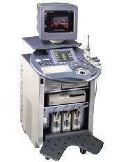
'GE is defining a new age of ultrasound. We call it Volume Ultrasound. GE's Voluson 730 Expert is a powerful system that enables real-time techniques for acquiring, navigating and analyzing volumetric images so that you can make clinical decisions with unprecedented confidence.'
Device Information and Specification
APPLICATIONS
Abdominal, breast, cardiac, musculoskeletal, neonatal, OB/GYN, pediatric, small parts, transcranial, urological, vascular
CONFIGURATION
15' high resolution non-interlaced flat CRT, 4 active probe ports
B-mode, M-mode, coded harmonic imaging (2-D), color flow mode (CFM), power Doppler imaging (PDI), color Doppler, pulsed wave Doppler, high pulse repetition frequency (HPRF) Doppler, tissue harmonic imaging, 3-D power Doppler
IMAGING OPTIONS
CrossXBeam spatial compounding, coded excitation , spatio-temporal image correlation (STIC), B-Flow (simultaneous imaging of tissue and blood flow), strain rate imaging (SRI)
OPTIONAL PACKAGE
STORAGE, CONNECTIVITY, OS
SonoView archiving and data management, network, HDD, DICOM 3.0, CD/DVD, MOD, USB, Windows-based
DATA PROCESSING
Digital beamformer with 512 system processing channel technology
H*W*D m (inch.)
1.43 * 0.69 * 1.02 (56 * 27 * 40)
WEIGHT
136 kg (300 lbs.)
Result Pages : |
Medical-Ultrasound-Imaging.com
former US-TIP.com
Member of SoftWays' Medical Imaging Group - MR-TIP • Radiology TIP • Medical-Ultrasound-Imaging
Copyright © 2008 - 2024 SoftWays. All rights reserved.
Terms of Use | Privacy Policy | Advertise With Us
former US-TIP.com
Member of SoftWays' Medical Imaging Group - MR-TIP • Radiology TIP • Medical-Ultrasound-Imaging
Copyright © 2008 - 2024 SoftWays. All rights reserved.
Terms of Use | Privacy Policy | Advertise With Us
[last update: 2023-11-06 01:42:00]


