Medical Ultrasound Imaging
Thursday, 9 May 2024
Info Sheets Out- side     | 'Harmonic B-Mode Imaging' Searchterm 'Harmonic B-Mode Imaging' found in 15 articles 1 term [ • ] - 2 definitions [• ] - 12 booleans [• ]Result Pages : • Harmonic B-Mode Imaging
Harmonic B-mode imaging takes advantage of the non-linear oscillation
of microbubbles. During harmonic imaging, the sound signal is transmitted at a frequency of around 1.5 to 2.0 MHz and received at twice this frequency. The microbubbles also reflect waves with wavelengths different from the transmitted one, the detectors can be set to receive only the latter ones and create only images of the contrast agent. Using bandpass filters the transmitted frequency is separated from the received signal to get improved visualization of vessels containing ultrasound contrast agents (USCAs). The signal to noise ratio during the presence of microbubbles in tissue is four- to fivefold higher at the harmonic compared with the basic frequency. Using harmonic B-mode imaging, harmonic frequencies produced by the ultrasound propagation through tissue have to be taken into account. The tissue reflection produces only a small amount harmonic energy compared to USCAs, but has to be removed by background subtraction for quantitative evaluation of myocardial perfusion. See also Non-linear Propagation. •
Also called B-mode echography, B-mode sonography, 2D-mode, and sonogram. B-mode ultrasound (Brightness-mode) is the display of a 2D-map of B-mode data, currently the most common form of ultrasound imaging. The development from A-mode to B-mode is that the ultrasound signal is used to produce various points whose brightness depends on the amplitude instead of the spiking vertical movements in the A-mode. Sweeping a narrow ultrasound beam through the area being examined while transmitting pulses and detecting echoes along closely spaced scan lines produces B-scan images. The vertical position of each bright dot is determined by the time delay from pulse transmission to return of the echo, and the horizontal position by the location of the receiving transducer element. To generate a rapid series of individual 2D images that show motion, the ultrasound beam is swept repeatedly. The returning sound pulses in B-mode have different shades of darkness depending on their intensities. The varying shades of gray reflect variations in the texture of internal organs. This form of display (solid areas appear white and fluid areas appear black) is also called gray scale. Different types of displayed B-mode images are: The probe movement can be performed manual (compound and static B-scanner) or automatic (real-time scanner). The image reconstruction can be parallel or sector type. See also B-Scan, 4B-Mode, and Harmonic B-Mode Imaging. Further Reading: News & More:
•
Harmonic imaging relies on detection of harmonics of the transmitted
frequency produced by bubble oscillation. This method is widely available on ultrasound scanners and uses the same array transducers as conventional imaging. A major limitation of the use of ultrasound contrast agents is the problem that signals from the microbubbles are mixed with those from tissue. Echoes from solid tissue and red blood cells are suppressed by harmonic imaging. In harmonic mode, the system transmits at one frequency, but is tuned to receive echoes preferentially at double that frequency, and the second harmonic echoes from the place of the bubble. Typically, the transmit frequency lies between 1.5 and 3 MHz and the receive frequency is selected by means of a bandpass filter whose center frequency lies between 3 and 6 MHz. Color Doppler and real-time harmonic spectral Doppler modes have also been implemented and show a level of tissue motion suppression not available in conventional modes. See also Harmonic B-Mode Imaging, and Harmonic Power Doppler. Further Reading: Basics:
News & More:
•  From GE Healthcare.;
'GE is defining a new age of ultrasound. We call it Volume Ultrasound. GE's Voluson 730 Expert is a powerful system that enables real-time techniques for acquiring, navigating and analyzing volumetric images so that you can make clinical decisions with unprecedented confidence.'
Device Information and Specification
APPLICATIONS
Abdominal, breast, cardiac, musculoskeletal, neonatal, OB/GYN, pediatric, small parts, transcranial, urological, vascular
CONFIGURATION
15' high resolution non-interlaced flat CRT, 4 active probe ports
B-mode, M-mode, coded harmonic imaging (2-D), color flow mode (CFM), power Doppler imaging (PDI), color Doppler, pulsed wave Doppler, high pulse repetition frequency (HPRF) Doppler, tissue harmonic imaging, 3-D power Doppler
IMAGING OPTIONS
CrossXBeam spatial compounding, coded excitation , spatio-temporal image correlation (STIC), B-Flow (simultaneous imaging of tissue and blood flow), strain rate imaging (SRI)
OPTIONAL PACKAGE
STORAGE, CONNECTIVITY, OS
SonoView archiving and data management, network, HDD, DICOM 3.0, CD/DVD, MOD, USB, Windows-based
DATA PROCESSING
Digital beamformer with 512 system processing channel technology
H*W*D m (inch.)
1.43 * 0.69 * 1.02 (56 * 27 * 40)
WEIGHT
136 kg (300 lbs.)
• 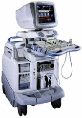 From GE Healthcare.;
'Vivid 7 Dimension, a premier cardiovascular ultrasound system from GE Healthcare, expands on the strength of a powerful imaging platform to offer new, innovative technology of dimensional proportions.'
Device Information and Specification
CONFIGURATION
Multi-frequency, linear, convex, phased, sector
B-mode, C-mode, M-mode (and 2-D), triplex mode, harmonic imaging, color flow mapping, 3D ultrasound display, power Doppler imaging (PDI), color Doppler, pulsed wave Doppler, continuous wave Doppler, tissue velocity imaging (TVI), tissue type imaging (TTI), strain rate imaging (SRI), tissue synchronization imaging (TSI)
IMAGING OPTIONS
CINE review with 5 speed types, bi- andtri-plane imaging with e.g. stress echo and tissue synchronization imaging
STORAGE, CONNECTIVITY, OS
Patient and image archive, HDD, MOD, DVD, USB flash card, DICOM 3.0 Windows-based
DATA PROCESSING
Digital beamformer with 1024 system processing channel technology
H*W*D m (inch.)
1.58 * 0.64 * 0.89 (62 * 25 * 35)
WEIGHT
191 kg (420 lbs.)
POWER CONSUMPTION
less than 2 KVA
Result Pages : | Share This Page Look Ups |
Medical-Ultrasound-Imaging.com
former US-TIP.com
Member of SoftWays' Medical Imaging Group - MR-TIP • Radiology TIP • Medical-Ultrasound-Imaging
Copyright © 2008 - 2024 SoftWays. All rights reserved.
Terms of Use | Privacy Policy | Advertise With Us
former US-TIP.com
Member of SoftWays' Medical Imaging Group - MR-TIP • Radiology TIP • Medical-Ultrasound-Imaging
Copyright © 2008 - 2024 SoftWays. All rights reserved.
Terms of Use | Privacy Policy | Advertise With Us
[last update: 2023-11-06 01:42:00]