Medical Ultrasound Imaging
Wednesday, 8 May 2024
Info Sheets Out- side     | 'Coded Excitation' Searchterm 'Coded Excitation' found in 5 articles 1 term [ • ] - 4 definitions [• ] Result Pages : • Coded Excitation
Increasing the frequency of the transmitted power improves the image quality of ultrasound, but the improvement in resolution results in a decreased signal to noise ratio (SNR). Higher acoustic power levels can prevent the loss in SNR, but among other reasons, ultrasound regulations limit this to avoid heating or cavitation. Coded excitation increase the signal to noise ratio without the loss of resolution by using coded waveforms. Coded excitation allows transmitting a long wide-band pulse with more acoustic power and high penetration of the sound beam. •
The perfect image quality is dependent on some assumptions of the propagation of ultrasound waves in tissues after generating in an imaging system. These assumptions are important for the developing of optimal ultrasound imaging systems.
•
The propagation of ultrasound is straight ahead.
•
•
•
•
The amplitudes of the echoes are proportional to the difference of the acoustical impedance caused by different tissue layers.
See also Coded Excitation, Validation and Refraction Artifact, Q-Value, Ultrasound Phantom, Dead Zone, Narrow Bandwidth. Further Reading: News & More: • 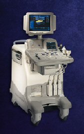 From GE Healthcare.;
Versatile High Performance System 'The LOGIQ 5 Expert ultrasound system delivers the premium performance advantage of TruScan architecture in a versatile high performance package. Advanced capabilities for outstanding clinical performance are available with the efficiency and productivity needed to meet clinical demands.'
Device Information and Specification
APPLICATIONS
Abdominal, cardiac, musculoskeletal, neonatal, OB/GYN, small parts, transcranial, urologic, vascular
CONFIGURATION
Normal system
B-mode, M-mode, anatomic M-mode creation and adjustment, triplex, 3D ultrasound, pulsed wave Doppler, continuous wave Doppler, power Doppler, color Doppler, spectral Doppler
IMAGING OPTIONS
OPTIONAL PACKAGE
STORAGE, CONNECTIVITY, OS
On-board patient, image and reporting archive, HDD, CD-ROM disk burner included, PCMCIA, USB, Windows-based OS
DATA PROCESSING
H*W*D m (inch.)
1.45 * 0.52 * 0.99 (57 * 20 * 39)
WEIGHT
180 kg (397 lbs.)
POWER CONSUMPTION
less than 1.5 KVA
•  From GE Healthcare.;
'GE is defining a new age of ultrasound. We call it Volume Ultrasound. GE's Voluson 730 Expert is a powerful system that enables real-time techniques for acquiring, navigating and analyzing volumetric images so that you can make clinical decisions with unprecedented confidence.'
Device Information and Specification
APPLICATIONS
Abdominal, breast, cardiac, musculoskeletal, neonatal, OB/GYN, pediatric, small parts, transcranial, urological, vascular
CONFIGURATION
15' high resolution non-interlaced flat CRT, 4 active probe ports
B-mode, M-mode, coded harmonic imaging (2-D), color flow mode (CFM), power Doppler imaging (PDI), color Doppler, pulsed wave Doppler, high pulse repetition frequency (HPRF) Doppler, tissue harmonic imaging, 3-D power Doppler
IMAGING OPTIONS
CrossXBeam spatial compounding, coded excitation , spatio-temporal image correlation (STIC), B-Flow (simultaneous imaging of tissue and blood flow), strain rate imaging (SRI)
OPTIONAL PACKAGE
STORAGE, CONNECTIVITY, OS
SonoView archiving and data management, network, HDD, DICOM 3.0, CD/DVD, MOD, USB, Windows-based
DATA PROCESSING
Digital beamformer with 512 system processing channel technology
H*W*D m (inch.)
1.43 * 0.69 * 1.02 (56 * 27 * 40)
WEIGHT
136 kg (300 lbs.)
•
The waveform is the record of a signal that varies over time. A blood flow signal for example, usually varies periodically with the cardiac cycle. See also Coded Excitation, and Pulse Volume Recording. Further Reading: News & More:
Result Pages : | Share This Page Look Ups |
Medical-Ultrasound-Imaging.com
former US-TIP.com
Member of SoftWays' Medical Imaging Group - MR-TIP • Radiology TIP • Medical-Ultrasound-Imaging
Copyright © 2008 - 2024 SoftWays. All rights reserved.
Terms of Use | Privacy Policy | Advertise With Us
former US-TIP.com
Member of SoftWays' Medical Imaging Group - MR-TIP • Radiology TIP • Medical-Ultrasound-Imaging
Copyright © 2008 - 2024 SoftWays. All rights reserved.
Terms of Use | Privacy Policy | Advertise With Us
[last update: 2023-11-06 01:42:00]