Medical Ultrasound Imaging
Wednesday, 8 May 2024
Info Sheets Out- side     | 'A-Scan' Searchterm 'A-Scan' found in 9 articles 1 term [ • ] - 8 definitions [• ] Result Pages : • A-Scan
A-scans are used in ophthalmologic scanning, to detect and monitor pregnancy problems, and screen intracranial mass lesions by using A-modes. A-scan ultrasound biometry, commonly referred to as an A-scan, is a routine diagnostic test used in ophthalmology. The A-scan provides data on the shape of the eye, which is a major determinant in common sight disorders. Ultrasound scanners used in this type of test require usually direct contact with the eye. See also A-Mode, Oculoplethysmography, Ultrasound Biomicroscopy, B-Scan, C-Scan and D-Scan. Further Reading: News & More:
•
A-mode (Amplitude-mode) ultrasound is a technique used to assess organ dimensions and determine the depth of an organ. While A-mode technology was previously employed in midline echoencephalography for rapid screening of intracranial mass lesions and ophthalmologic scanning, it is now considered obsolete in medical imaging. Nonetheless, the A-mode scan has found applications in early pregnancy assessment (specifically the detection of fetal heartbeats), cephalometry, and placental localization.
When the ultrasound beam encounters an anatomic boundary, the received sound impulse is processed to appear as a vertical reflection of a point. On the display, it looks like spikes of different heights (the amplitude). The intensity of the returning impulse determined the height of the vertical reflection and the time it took for the impulse to make the round trip would determine the space between verticals. The distance between these spikes can be measured accurately by dividing the speed of sound in tissue (1540 m/sec) by half the sound travel time. During an echoencephalography scan, the first A-mode scan is acquired from the right side of the head and captured on film. Subsequently, the probe is positioned at the corresponding point on the left side, and a second exposure is captured on the same film, displaying inverted spikes. The A-mode ultrasound could be used to identify structures normally located in the midline of the brain such as the third ventricle and falx cerebri. The midline structures would be aligned in normal patients but show displacement in patients with mass lesion such as a subdural, epidural, or intracranial hemorrhage. See also 2D Ultrasound, 3D Ultrasound, 4D Ultrasound, Ultrasound Biomicroscopy, A-scan, B-mode and the Infosheet about ultrasound modes. • 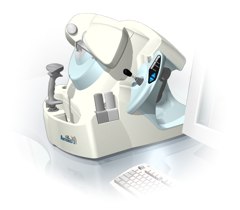 From Ultralink LLC;
'Artemis is a very high frequency (VHF) ultrasound eye scanner. In use, the patient leans forward placing their head onto an adjustable headrest. The headrest's unique design permits the patient to pull away quickly from the scanner if desired. An eyecup filled with a saline-based interface fluid couples the ultrasound signal to the eye, while a precision mechanism moves the transducer past the front of the eye. During the accurately controlled arc motion of the transducer, which lasts less than one second, many thousands of ultrasound samples are digitized. Following a scan, signal analysis is performed on a PC-compatible microcomputer, and the data are available for immediate viewing on an LCD monitor or disk storage. Artemis is very flexible; many adjustments to the scanning parameters are possible to customize the scan to your clinical needs. Functions are provided for centering the scan about the optical axis of the eye. The starting location of the scans as well as the extent can be varied as desired, to view image planes through the eye at different angles.' See also Ultrasound Biomicroscopy, A-Mode and A-Scan. Further Reading: News & More:
•
B-scan ultrasonography, or B-scan, is a diagnostic test, for example used in ophthalmology to produce a two-dimensional, cross-sectional view of the eye and the orbit. The presentation of the reflected pulses are displayed in rectangular coordinates, in which the travel time of an ultrasound pulse is represented as a shift along one axis, and the probe movement is represented as a shift along the other axis. See also Ultrasound Imaging Procedures, A-Scan, C-Scan and D-Scan. •
B-scan comined with D-scan (D=Depth) is used to avoid image inhomogeneity. Different transmitter signals for each depth are applied and prefiltered pseudoinversely according to the transfer properties of the covering tissue. Pulse compression techniques with nonlinearly frequency modulated signals are used to gain the required energy for inverse filtering. D-scan is a modified C-scan used in nondestructive testing with the display of amplitudes. In the 2D graphical presentation, time of flight values are displayed in the top view on a test surface. See also A-Scan, B-Scan and C-Scan. Result Pages : | Share This Page Look Ups |
Medical-Ultrasound-Imaging.com
former US-TIP.com
Member of SoftWays' Medical Imaging Group - MR-TIP • Radiology TIP • Medical-Ultrasound-Imaging
Copyright © 2008 - 2024 SoftWays. All rights reserved.
Terms of Use | Privacy Policy | Advertise With Us
former US-TIP.com
Member of SoftWays' Medical Imaging Group - MR-TIP • Radiology TIP • Medical-Ultrasound-Imaging
Copyright © 2008 - 2024 SoftWays. All rights reserved.
Terms of Use | Privacy Policy | Advertise With Us
[last update: 2023-11-06 01:42:00]