Medical Ultrasound Imaging
Wednesday, 8 May 2024
Info Sheets Out- side     | 'Contrast Imaging Techniques' Searchterm 'Contrast Imaging Techniques' found in 17 articles 1 term [ • ] - 2 definitions [• ] - 14 booleans [• ]Result Pages : • Contrast Imaging Techniques
Many different contrast imaging techniques have been developed. Most are either variations, hybrids, or combinations of the following ultrasound techniques: See also Coherent Contrast Imaging, Ultrasound Picture and Targeted Contrast Imaging. Further Reading: Basics:
•
Bubble specific imaging methods rely usually on non-linear imaging modes. These contrast imaging techniques are designed to suppress the echo from tissue in relation to that from a microbubble contrast agent. Stimulated acoustic emission (SAE) and phase / pulse inversion imaging mode (PIM) are bubble specific modes, which can image the tissue specific phase. In SAE mode bubble rupture is seen as a transient bright signal in B-mode and as a characteristic mosaic-like effect in velocity 2D color Doppler. PIM are Doppler modes and detect non-linear echoes from microbubbles. In pulse inversion imaging modes the transducer bandwidth extends, resulting in improved spatial resolution and more contrast. See also Contrast Pulse Sequencing, Microbubble Scanner Modification, Narrow Bandwidth, Contrast Medium, Dead Zone. Further Reading: Basics:
•
(CEUS) Contrast agents increase the reflection of ultrasonic energy, improve the signal to noise ratio and caused by that the detection of abnormal microvascular and macrovascular disorders. Contrast enhanced ultrasound is used in abdominal ultrasound (liver sonography) as well as in cerebrovascular examinations e.g., for an accurate grading of carotid stenosis. The used contrast agents are safe and well tolerated. The quality of the enhancement depends on:
•
the concentration of the contrast agent;
•
the type of injection, flow rate;
•
the patient characteristics;
•
the microbubble quality and properties of the filling gas and the shell.
The additional use of ultrasound contrast agents (USCAs) may overcome typical limitations like poor contrast of B-mode imaging or limited sensitivity of Doppler techniques. The development of new ultrasound applications (e.g., blood flow imaging, perfusion quantification) depends also from the development of pulse sequences for bubble specific imaging. In addition, contrast enhanced ultrasound improves the monitoring of ultrasound guided interventions like RF thermal ablation. See also Contrast Enhanced Doppler Imaging, Contrast Harmonic Imaging, Contrast Imaging Techniques and Contrast Pulse Sequencing. Further Reading: News & More:
•  From Philips Medical Systems;
'The SONOS 5500 has all the advanced features you want in a high-performance ultrasound system. Exceptional image quality, revolutionary contrast imaging tools, unique quantitative techniques, extensive applications, and an intuitive user interface allow the most comprehensive echo exams possible.' Specifications for this system will be available soon. •  From GE Healthcare.;
'GE is defining a new age of ultrasound. We call it Volume Ultrasound. GE's Voluson 730 Expert is a powerful system that enables real-time techniques for acquiring, navigating and analyzing volumetric images so that you can make clinical decisions with unprecedented confidence.'
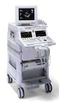
Device Information and Specification
APPLICATIONS
Abdominal, breast, cardiac, musculoskeletal, neonatal, OB/GYN, pediatric, small parts, transcranial, urological, vascular
CONFIGURATION
15' high resolution non-interlaced flat CRT, 4 active probe ports
B-mode, M-mode, coded harmonic imaging (2-D), color flow mode (CFM), power Doppler imaging (PDI), color Doppler, pulsed wave Doppler, high pulse repetition frequency (HPRF) Doppler, tissue harmonic imaging, 3-D power Doppler
IMAGING OPTIONS
CrossXBeam spatial compounding, coded excitation , spatio-temporal image correlation (STIC), B-Flow (simultaneous imaging of tissue and blood flow), strain rate imaging (SRI)
OPTIONAL PACKAGE
STORAGE, CONNECTIVITY, OS
SonoView archiving and data management, network, HDD, DICOM 3.0, CD/DVD, MOD, USB, Windows-based
DATA PROCESSING
Digital beamformer with 512 system processing channel technology
H*W*D m (inch.)
1.43 * 0.69 * 1.02 (56 * 27 * 40)
WEIGHT
136 kg (300 lbs.)
Result Pages : | Share This Page Look Ups |
Medical-Ultrasound-Imaging.com
former US-TIP.com
Member of SoftWays' Medical Imaging Group - MR-TIP • Radiology TIP • Medical-Ultrasound-Imaging
Copyright © 2008 - 2024 SoftWays. All rights reserved.
Terms of Use | Privacy Policy | Advertise With Us
former US-TIP.com
Member of SoftWays' Medical Imaging Group - MR-TIP • Radiology TIP • Medical-Ultrasound-Imaging
Copyright © 2008 - 2024 SoftWays. All rights reserved.
Terms of Use | Privacy Policy | Advertise With Us
[last update: 2023-11-06 01:42:00]