Medical Ultrasound Imaging
Tuesday, 14 May 2024
Info Sheets Out- side     | 'Angiography' p2 Searchterm 'Angiography' found in 9 articles 1 term [ • ] - 8 definitions [• ] Result Pages : •
(MIP) Angiography (Doppler) images can be processed by Maximum Intensity Projection to interactively create different projections. Although the maximum intensity projection (MIP) post processing algorithm is sensitive to high signal from inflowing spins as used in MRI, it is also sensitive to high signal of any other etiology as used in ultrasound imaging. The MIP connects the high intensity dots of the blood vessels in three dimensions, providing an angiogram that can be viewed from any projection. Each point in the MIP represents the highest intensity experienced in that location on any partition within the imaging volume. For complete interpretation the base slices should also be reviewed individually and with multiplanar reconstruction (MPR) software. The MIP can then be displayed in a CINE format or filmed as multiple images acquired from different projections. See also 3D Ultrasound. •
(OPG) The Oculoplethysmography is used to detect a hemodynamically significant stenosis of the carotid circulation, by measuring the arrival times of pulses in the eye and ear. Indirectly the blood flow in the ophthalmic artery is measured, which branches off the carotid artery and supplies blood to the eye. A delay between pulse arrival in the eye and ear is an indication of a hemodynamically significant stenosis in the vessel whose corresponding pulse is delayed. Other ultrasound or angiography procedures are necessary to confirm the diagnosis and have also replaced this technique. See also Plethysmography and Ultrasound Biomicroscopy. Further Reading: News & More:
• 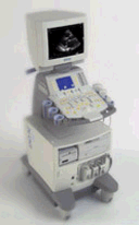 From Siemens Medical Systems;
'This high-performance, multi-specialty system supports and improves your daily ultrasound routine. Embedded DICOM creates the integrated foundation for a complete connectivity solution while MultiHertz™ multiple frequency imaging and Tissue Harmonic Imaging (THI) expands the clinical versatility of the system. Advanced applications such as stress echo, SieScape™ panoramic imaging, and transesophageal imaging can be seamlessly integrated.'
Device Information and Specification
CLINICAL APPLICATION
Widest range of applications
CONFIGURATION
Compact, mobile system
Wideband MultiHertz™ multiple frequency
IMAGING OPTIONS
OPTIONAL PACKAGE
Software upgradeability to advanced clinical application
IMAGING ENHANCEMENTS
Precision MotionCapture, Synthetic aperture technology
STORAGE
Patient and image database management system
DATA PROCESSING
Parallel and quad signal processing
WEIGHT
Lightweight
• In September 2000 Siemens Medical Engineering Group, bought Acuson Corporation (Mountain View, California) for approximately $700 million. Ultrasound Systems: Ultrasound Systems (older):
Contact Information
Result Pages : | Share This Page Look Ups |
Medical-Ultrasound-Imaging.com
former US-TIP.com
Member of SoftWays' Medical Imaging Group - MR-TIP • Radiology TIP • Medical-Ultrasound-Imaging
Copyright © 2008 - 2024 SoftWays. All rights reserved.
Terms of Use | Privacy Policy | Advertise With Us
former US-TIP.com
Member of SoftWays' Medical Imaging Group - MR-TIP • Radiology TIP • Medical-Ultrasound-Imaging
Copyright © 2008 - 2024 SoftWays. All rights reserved.
Terms of Use | Privacy Policy | Advertise With Us
[last update: 2023-11-06 01:42:00]