Medical Ultrasound Imaging
Monday, 20 May 2024
Info Sheets Out- side     | 'Image Quality' p7 Searchterm 'Image Quality' found in 40 articles 1 term [ • ] - 39 definitions [• ] Result Pages : • 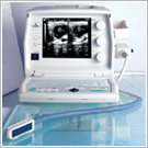 From Medison Co.,Ltd.;
'SONOVET 600 and Veterinarian SONOVET 600 provides an improved image quality for accurate and reliable diagnosis for bovine and equine reproductive organ scanning, and small animal general abdominal examination. For higher resolution imaging, the industry standard linear and convex probes are available with the extensive measurement function.' • 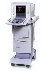 From Kontron Medical SAS;
'The Sigma 330 is a versatile, digital, mobile Ultrasound System, upgradeable from 2D to Doppler, Color Flow Mapping and 3D. The Sigma 330 displays excellent image quality and superb Doppler and CFM sensitivity, employing ATEC™ (Advanced Tissue Echo Cancellation), a revolutionary technology developed by the Kontron Medical R&D Center, which removes tissue artifacts ('ghosting').' Specifications for this system will be available soon. • The field of medical imaging offers numerous career opportunities, and one profession is that of a sonographer. Sonographers play a critical role in healthcare by utilizing ultrasound technology to create images of the body's internal structures. •
Becoming a Sonographer: The educational and professional requirements for sonographers can vary from country to country. The duration of these programs can range from one to four years, depending on the country and level of qualification. The typical path in the United States begins with obtaining a post-secondary education in diagnostic medical sonography from an accredited program. These programs usually result in an associate's or bachelor's degree. Coursework typically covers anatomy, physiology, medical ethics, ultrasound physics, and specialized sonography techniques. Additionally, students gain practical experience through clinical internships in healthcare facilities. After completing their education, aspiring sonographers can choose to obtain professional certification through organizations such as the American Registry for Diagnostic Medical Sonography (ARDMS) or the American Registry of Radiologic Technologists (ARRT). Certification often requires passing examinations that assess knowledge and competency in specific areas of sonography. Many countries also have certification or registration requirements for sonographers. These certifications are typically obtained through professional bodies or organizations specific to each country. Examples include the Canadian Association of Registered Diagnostic Ultrasound Professionals (CARDUP) in Canada, the Australian Sonographers Accreditation Registry (ASAR) in Australia, and the Society and College of Radiographers (SCoR) in the United Kingdom. •
Job Description: Sonographers are skilled professionals who operate ultrasound machines and perform sonograms on patients. They work closely with physicians and other healthcare professionals to provide accurate and high-quality diagnostic images. Using sound waves, sonographers capture images of organs, tissues, and blood flow patterns, which are then used by medical practitioners to diagnose and monitor various medical conditions. Sonographers must have a comprehensive understanding of anatomy, physiology, and sonographic techniques to optimize image quality. They interact directly with patients, explaining procedures, addressing concerns, and ensuring patient comfort throughout the scanning process. Documentation of findings and communication with the medical team are also essential responsibilities. Some aspect of the job can be demanding, while sonographers often spend long hours on their feet, positioning and maneuvering patients during scans. Dealing with patients who are in pain, anxious, or difficult to scan requires empathy, patience, and excellent interpersonal skills. Sonographers often work in fast-paced environments, juggling multiple patients and procedures throughout the day. Effective time management is essential to ensure that scans are performed efficiently without compromising quality. Adhering to schedules and meeting the demands of the healthcare facility can add to the workload and stress levels. •
Salary Outlook: The salary of a sonographer can vary, based on factors such as experience, specialization, geographic location, and work setting. According to the U.S. Bureau of Labor Statistics, as of May 2021, the median annual wage for diagnostic medical sonographers was $77,740. Sonographers working in specialized hospitals, outpatient care centers, and diagnostic imaging centers tend to earn higher salaries compared to those in physician offices or government facilities. The salary prospects for sonographers outside the United States can vary significantly based on factors such as the country's economic conditions, healthcare system, demand for sonographers, and cost of living. •
Future Outlook: The future outlook for sonographers appears highly favorable. The demand for ultrasound imaging continues to grow due to advancements in medical technology and an aging population. This increasing demand for sonographers is expected to result in good job prospects and potential career advancement opportunities. Monitoring job markets, understanding regulatory requirements, and networking with professionals in international healthcare communities can provide valuable insights into future opportunities. See also Handheld Ultrasound, Ultrasound Machine, Sonography, Portable Ultrasound Machine, Ultrasound Accessories and Supplies, Environmental Protection and Ultrasound Technology. •
Common ultrasound supplies that are often used in conjunction with ultrasound imaging:
•
Ultrasound Gel: A water-based gel used as a coupling agent between the transducer and the patient's skin. It helps eliminate air pockets and ensures good sound wave transmission. •
Probe Covers: Disposable covers designed to maintain hygiene and prevent cross-contamination. These covers are placed over the transducer before each examination. •
Cleaning Wipes: Alcohol-based or disinfectant wipes used for cleaning and disinfecting the transducer and other equipment surfaces. Specific cleaning solutions are recommended by the ultrasound equipment manufacturer for thorough cleaning of transducers. •
Gel Warmers: Devices used to warm ultrasound gel, providing patient comfort during the examination. •
Needle Guides: Attachments or brackets that assist in accurate needle placement during ultrasound-guided procedures such as biopsies or injections. •
Positioning Aids: Cushions, wedges, or straps designed to help position patients correctly and comfortably during ultrasound exams. Common ultrasound accessories that are often used in conjunction with ultrasound imaging: •
Transducer Storage Rack: A dedicated rack or holder to store transducers safely when not in use, helping to prevent damage. •
Storage and Archiving Solutions: External hard drives, network storage, or cloud-based systems for long-term storage and backup of ultrasound images and reports. Possibly specialized printers that produce hard copies of ultrasound images for immediate documentation and patient records. •
Power Supply and Transducer Cable Extenders: Extension cables used to increase the length of transducer cables for more flexibility during examinations. Adequate power sources or uninterrupted power supply (UPS) to ensure continuous operation of the ultrasound machine during power outages or fluctuations. •
Reporting Templates and Software: Customizable reporting templates and software solutions that facilitate efficient and standardized reporting of ultrasound findings. •
Phantom Devices: Artificial tissue-like structures or phantoms used for training, calibration, and quality assurance purposes to evaluate image quality and system performance. Consult with ultrasound equipment vendors or professionals in the field to determine the specific accessories and supplies that best suit your imaging needs and specialty. See also Equipment Preparation, Environmental Protection, Portable Ultrasound Machine, Ultrasound Technology, Ultrasound System Performance and Sonographer. •
(UCA / USCA) Ultrasonography is the most commonly performed diagnostic imaging procedure. The introduction of sonographic contrast media into routine practice modifies the use of ultrasound in a variety of clinical applications. USCAs consist of microbubbles filled with air or gases and can be classified according to their pharmacokinetics. Among the blood pool agents, transpulmonary ultrasound contrast agents offer higher diagnostic potential compared to agents that cannot pass the pulmonary capillary bed after a peripheral intravenous injection. In addition to their vascular phase, some USCAs can exhibit a tissue- or organ-specific phase. The sonogram image quality is improved either by decreasing the reflectivity of the undesired interfaces or by increasing the backscattered echoes from the desired regions. Different types of ultrasound contrast agents: Ultrasound contrast agents act as echo-enhancers, because of the high different acoustic impedance at the interface between gas and blood. The enhanced echo intensity is proportional to the change in acoustical impedance as the sound beam crosses from the blood to the gas in the bubbles. The ideal qualities of an ultrasound contrast agent:
•
high echogenicity;
•
low attenuation;
•
low blood solubility;
•
low diffusivity;
•
ability to pass through the pulmonary capillary bed;
•
lack of biological effects with repeat doses.
A typical ultrasound contrast agent consists of a thin flexible or rigid shell composed of albumin, lipid, or polymer confining a gas such as nitrogen, or a perfluorocarbon. The choice of the microbubble shell and gas has an important influence on the properties of the agent. Current generations of microbubbles have a diameter from 1 μm to 5 μm. The success of these agents is mostly dependent on the small size and on the stability of their shell, which allows passage of the microbubbles through the pulmonary circulation. Microbubbles must be made smaller than the diameter of capillaries or they would embolize and be ineffective and perhaps even dangerous. The reflectivity of these microbubbles is proportional to the fourth power of a particle diameter but also directly proportional to the concentration of the contrast agent particles themselves. Ultrasound contrast agents produce unique acoustic signatures that allow to separate their signal from tissue echoes and to depict whether they are moving or stationary. This enables the detection of capillary flow and of targeted microbubbles that are retained in tissues such as normal liver. The new generation of contrast media is characterized by prolonged persistence in the vascular bed which provides consistent enhancement of the arterial Doppler signal. Contrast agents make it also possible to perform dynamic and perfusion studies. Targeted contrast imaging agents are for example taken up by the phagocytic cell systems and thus have liver/spleen specific effects. See also Ultrasound Contrast Agent Safety, Adverse Reaction, Tissue-Specific Ultrasound Contrast Agent, and Bubble Specific Imaging. Further Reading: Basics:
News & More:
Result Pages : | Share This Page Look Ups |
Medical-Ultrasound-Imaging.com
former US-TIP.com
Member of SoftWays' Medical Imaging Group - MR-TIP • Radiology TIP • Medical-Ultrasound-Imaging
Copyright © 2008 - 2024 SoftWays. All rights reserved.
Terms of Use | Privacy Policy | Advertise With Us
former US-TIP.com
Member of SoftWays' Medical Imaging Group - MR-TIP • Radiology TIP • Medical-Ultrasound-Imaging
Copyright © 2008 - 2024 SoftWays. All rights reserved.
Terms of Use | Privacy Policy | Advertise With Us
[last update: 2023-11-06 01:42:00]