Medical Ultrasound Imaging
Wednesday, 8 May 2024
Info Sheets Out- side     | 'Superior' p2 Searchterm 'Superior' found in 11 articles 1 term [ • ] - 10 definitions [• ] Result Pages : •
If available, some graphic aids can be helpful to show image orientations. 1) A graphic icon of the labeled primary axes (A, L, H) with relative lengths given by direction sines and orientation as if viewed from the normal to the image plane can help orient the viewer, both to identify image plane orientation and to indicate possible in plane rotation. 2) Ingraphic prescription of obliques from other images, a sample original image with an overlaid line or set of lines indicating the intersection of the original and oblique image planes can help orient the viewer.
•
•
In all cases the scanning surface is assigned to the top of the image. The orientation of single oblique slices can be specified by rotating a slice in one of the basic orientations toward one of the other two basic orthogonal planes about an axis defined by the intersection of the 2 planes. See also Histogram. • 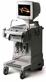 From Medison Co.,Ltd.;
'It's the dawn of a new Evolution in affordable digital color ultrasound At Medison, we have a history of innovation. We were the first to make the leap to the third dimension when we introduced our groundbreaking digital 3D ultrasound platform with Live 3D™ technology in 1998, making it possible to capture true 3D volume data in real-time. Today, we're proud to introduce the SONOACE , the world's first true 3D color ultrasound system designed to bring the power of real-time 3D imaging to women's health specialists at a breakthrough price.' 'SONOACE 8000 Live PRIME, the true 3D ultrasound system designed to bring the power of live 3D in your hands. SONOACE 8000 Live PRIME offers superior image quality thanks to our new C-Square Technology and newly applied PSAD Beamformer.' • 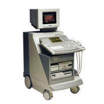 From Siemens Medical Systems;
'The SONOLINE Elegra™ ultrasound system's proven track record in connectivity, ergonomic design, and strong line of transducers make this system a top choice for the most demanding radiology department. The unparalleled union of real-time image processing and superior image quality creates a powerful opportunity for significant increases in diagnostic confidence.' Specifications for this system will be available soon. •  From Medison Co.,Ltd.;
'Putting the Power of Portable Ultrasound in Veterinarians' Hands Personal Veterinary Digital Ultrasound Yes, SONOVET2000 is good news for our four-legged friends, veterinarians and animal husbandry professionals. Portable and wearable, it puts the power of digital ultrasound scanning in your hands for non-invasive diagnosis anywhere, anytime. With it's on-site capabilities, SONOVET2000 raises the bar for animal health care quality and your working efficiency. And it comes with the superior imaging power of Medison's all-digital beamforming technology. Enjoy a higher productivity and efficiency at a surprisingly competitive price.' •
(TEE) Transesophageal echocardiography provides a superior view of cardiac anatomy compared with transthoracic echocardiography. TEE is performed by the introduction of a probe attached to a fiberoptic endoscope into the esophagus. Caused by the position close to the heart e.g., clot finding and the view of the mitral valve are improved. Indications:
•
aortic atherosclerotic disease;
•
aortic dissection;
•
artificial mitral valves;
•
clots inside the left atrium;
•
cardiac infections;
•
masses or clots in the heart.
The piezoelectric crystal creating the acoustic power is mounted on the gastroscope that must be swallowed by the patient. This endoscopic transducer is miniaturized to approximately the size of a fingernail. Usually the probe is in place for an average of 15 minutes, to numb the surface a topical anesthetic is sprayed into the throat, in addition a conscious sedation is recommended. See also Myocardial Contrast Echocardiography, Stress Echocardiogram, M-Mode Echocardiography, Contrast Enhanced Ultrasound and Vascular Ultrasound Contrast Agents. Result Pages : | Share This Page Look Ups |
Medical-Ultrasound-Imaging.com
former US-TIP.com
Member of SoftWays' Medical Imaging Group - MR-TIP • Radiology TIP • Medical-Ultrasound-Imaging
Copyright © 2008 - 2024 SoftWays. All rights reserved.
Terms of Use | Privacy Policy | Advertise With Us
former US-TIP.com
Member of SoftWays' Medical Imaging Group - MR-TIP • Radiology TIP • Medical-Ultrasound-Imaging
Copyright © 2008 - 2024 SoftWays. All rights reserved.
Terms of Use | Privacy Policy | Advertise With Us
[last update: 2023-11-06 01:42:00]