Medical Ultrasound Imaging
Saturday, 27 April 2024
Info Sheets Out- side     | '3D Ultrasound' Searchterm '3D Ultrasound' found in 23 articles 1 term [ • ] - 18 definitions [• ] - 4 booleans [• ]Result Pages : • 3D Ultrasound
In 3D ultrasound (US) several 2D images are acquired by moving the probe across the body surface or rotating inserted probes. 3D-mode uses the same basic concept of a 2D ultrasound but rather than take the image from a single angle, the sonographer takes a volume image. The volume image that is displayed on the screen is a software rendering of all of the detected soft-tissue combined by specialized computer software to form three-dimensional images. The 3D volume rendering technique (VR) does not rely on segmentation (segmentation techniques are difficult to apply to ultrasound pictures) and makes it possible to obtain clear 3D ultrasound images for clinical diagnosis. A 3D ultrasound produces a still image. Diagnostic US systems with 3D display functions and linear array probes are mainly used for obstetric and abdominal applications. The combination of contrast agents, harmonic imaging and power Doppler greatly improves 3D US reconstructions. 3D imaging shows a better look at the organ being examined and is used for:
•
Detection of abnormal fetus development, e.g. of the face and limbs.
•
Visualization of e.g. the colon and rectum.
•
Pictures of blood flow in various organs or a fetus.
Fusion 3D imaging methods for generating compound images from two sets of ultrasound images (B-mode and Doppler images) enable the observation of the structural relationships between lesions and their associated blood vessels in three dimensions (maximum intensity projection). Further Reading: News & More:
•
As far as ultrasound is concerned, 4D ultrasound (also referred to as live 3D ultrasound or 4B-mode) is the latest ultrasound technology - the fourth dimension means length, width, and depth over time. 4D Ultrasound takes 3D ultrasound images and adds the element of time to the progress so that a moving three-dimensional image is seen on the monitor. A 4D scan takes the same amounts of time as a 2D or 3D scan; the difference is the ultrasound equipment being used. One advantage of a 4D fetal ultrasound to a 2D-mode is that parents can see how their baby will generally look like. However, there are different opinions over the medical advantages. To scan a 3D ultrasound image, the probe is swept over the maternal abdomen. A computer takes multiple images and renders the 3D picture. With 4D imaging, the computer takes the images as multiple pictures while the probe is hold still and a 3D image is simultaneously rendered in real time on a monitor. In most cases, the standard 2D ultrasound is taken, and then the 3D/4D scan capability is added if an abnormality is detected or suspected. The 3D/4D sonogram is then focused on a specific area, to provide the details needed to assess and diagnose a suspected problem. A quick 4D scan of the face of the fetus may be performed at the end of a routine exam, providing the parents with a photo. •
As far as ultrasound is concerned, 4D ultrasound (also referred to as live 3D ultrasound or 4B-mode) is the latest ultrasound technology - the fourth dimension means length, width, and depth over time. 4D Ultrasound takes 3D ultrasound images and adds the element of time to the progress so that a moving three-dimensional image is seen on the monitor. A 4D scan takes the same amounts of time as a 2D or 3D scan; the difference is the ultrasound equipment being used. One advantage of a 4D fetal ultrasound to a 2D-mode is that parents can see how their baby will generally look like. However, there are different opinions over the medical advantages. To scan a 3D ultrasound image, the probe is swept over the maternal abdomen. A computer takes multiple images and renders the 3D picture. With 4D imaging, the computer takes the images as multiple pictures while the probe is hold still and a 3D image is simultaneously rendered in real time on a monitor. In most cases, the standard 2D ultrasound is taken, and then the 3D/4D scan capability is added if an abnormality is detected or suspected. The 3D/4D sonogram is then focused on a specific area, to provide the details needed to assess and diagnose a suspected problem. A quick 4D scan of the face of the fetus may be performed at the end of a routine exam, providing the parents with a photo. See also Obstetric and Gynecologic Ultrasound, Pregnancy Ultrasound, Fetal Ultrasound and Abdominal Ultrasound. • 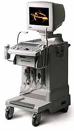 From Medison Co.,Ltd.;
'It's the dawn of a new Evolution in affordable digital color ultrasound At Medison, we have a history of innovation. We were the first to make the leap to the third dimension when we introduced our groundbreaking digital 3D ultrasound platform with Live 3D™ technology in 1998, making it possible to capture true 3D volume data in real-time. Today, we're proud to introduce the SONOACE , the world's first true 3D color ultrasound system designed to bring the power of real-time 3D imaging to women's health specialists at a breakthrough price.' 'SONOACE 8000 Live PRIME, the true 3D ultrasound system designed to bring the power of live 3D in your hands. SONOACE 8000 Live PRIME offers superior image quality thanks to our new C-Square Technology and newly applied PSAD Beamformer.' •
2D ultrasound imaging is a widely used technique in medical imaging that provides two-dimensional visual representations of internal structures. A handheld device known as a probe or transducer contains piezoelectric crystals that emit and receive ultrasound waves which penetrate tissues and bounce back as echoes. The echoes are detected and converted into electrical signals. These signals are processed and displayed on a monitor, creating a real-time 2D grayscale image, with different shades of gray representing various tissue densities. The brighter areas on the image correspond to structures that reflect more ultrasound waves, while darker areas represent structures that reflect fewer waves or are attenuated by intervening tissues. The 2D-mode (or B-mode) provides cross-sectional views of the scanned area, showing a single plane or slice of the scanned area at a time. Key Features and Uses of 2D Ultrasound: •
•
2D ultrasound is excellent for visualizing anatomical structures and detecting anomalies. It is widely used in obstetrics, gynecology, abdominal imaging and vascular examinations.
•
Due to its real-time capabilities, 2D ultrasound is utilized to guide various procedures, including biopsies, injections, and catheter insertions.
•
2D sonography can incorporate Doppler technology to assess blood flow in vessels, aiding in the diagnosis of vascular conditions and evaluating fetal circulation.
Comparison with 3D and 4D Ultrasound: •
Unlike 2D ultrasound, which generates a series of 2D images, 3D ultrasound creates a three-dimensional volume of the scanned area. This allows for more detailed visualization of complex structures, such as fetal facial features or organ morphology.
•
4D ultrasound adds the dimension of time to 3D imaging, resulting in dynamic three-dimensional videos. It enables the visualization of fetal movements and provides a more immersive experience. However, a 4D sonogram is not typically used for diagnostic purposes and is often employed in baby ultrasound examinations for bonding and enjoyment purposes.
See also Ultrasound Technology, Sonographer, Ultrasound Elastography, Obstetric and Gynecologic Ultrasound. Result Pages : | Share This Page Look Ups |
Medical-Ultrasound-Imaging.com
former US-TIP.com
Member of SoftWays' Medical Imaging Group - MR-TIP • Radiology TIP • Medical-Ultrasound-Imaging
Copyright © 2008 - 2024 SoftWays. All rights reserved.
Terms of Use | Privacy Policy | Advertise With Us
former US-TIP.com
Member of SoftWays' Medical Imaging Group - MR-TIP • Radiology TIP • Medical-Ultrasound-Imaging
Copyright © 2008 - 2024 SoftWays. All rights reserved.
Terms of Use | Privacy Policy | Advertise With Us
[last update: 2023-11-06 01:42:00]