Medical Ultrasound Imaging
Sunday, 19 May 2024
Info Sheets Out- side     | 'Handheld Ultrasound' p3 Searchterm 'Handheld Ultrasound' found in 17 articles 1 term [ • ] - 9 definitions [• ] - 7 booleans [• ]Result Pages : •
A transducer assembly is the configuration of an ultrasound transducer. Electronic array transducers are composed of multiple crystal elements e.g., linear array and phased array transducer assemblies. A handheld mechanical probe may contain a mechanically driven single element transducer, or a rotating wheel transducer. Static B-scanner (obsolete) had a cylindrical probe with a single disk-shaped piezoelectric crystal. Some probes are designed to be inserted in body cavities (vagina, rectum, esophagus) so that they can get closer to the organ being examined (uterus, prostate gland, heart); getting closer to the region of interest allow for a more detailed view. See also Endocavitary Echography, Endoscopic Ultrasound, Vaginal Transducer and Rectal Probe. •
A vaginal probe is a small ultrasound transducer (e.g. a biplane probe) that is inserted directly into the vagina. A sterile cover is slipped over the handheld transducer (also handle or probe), which is then covered with lubricating gel and placed in the vagina. This technique produces a sharp image, not only because of the close proximity to the uterus, but also because this transducer is more sensitive. •
2D ultrasound imaging is a widely used technique in medical imaging that provides two-dimensional visual representations of internal structures. A handheld device known as a probe or transducer contains piezoelectric crystals that emit and receive ultrasound waves which penetrate tissues and bounce back as echoes. The echoes are detected and converted into electrical signals. These signals are processed and displayed on a monitor, creating a real-time 2D grayscale image, with different shades of gray representing various tissue densities. The brighter areas on the image correspond to structures that reflect more ultrasound waves, while darker areas represent structures that reflect fewer waves or are attenuated by intervening tissues. The 2D-mode (or B-mode) provides cross-sectional views of the scanned area, showing a single plane or slice of the scanned area at a time. Key Features and Uses of 2D Ultrasound: •
•
2D ultrasound is excellent for visualizing anatomical structures and detecting anomalies. It is widely used in obstetrics, gynecology, abdominal imaging and vascular examinations.
•
Due to its real-time capabilities, 2D ultrasound is utilized to guide various procedures, including biopsies, injections, and catheter insertions.
•
2D sonography can incorporate Doppler technology to assess blood flow in vessels, aiding in the diagnosis of vascular conditions and evaluating fetal circulation.
Comparison with 3D and 4D Ultrasound: •
Unlike 2D ultrasound, which generates a series of 2D images, 3D ultrasound creates a three-dimensional volume of the scanned area. This allows for more detailed visualization of complex structures, such as fetal facial features or organ morphology.
•
4D ultrasound adds the dimension of time to 3D imaging, resulting in dynamic three-dimensional videos. It enables the visualization of fetal movements and provides a more immersive experience. However, a 4D sonogram is not typically used for diagnostic purposes and is often employed in baby ultrasound examinations for bonding and enjoyment purposes.
See also Ultrasound Technology, Sonographer, Ultrasound Elastography, Obstetric and Gynecologic Ultrasound. • 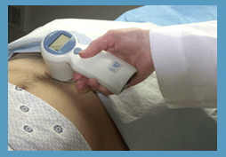 From Verathon Inc.;
'The BladderScan® BVI 6100 is a handheld, noninvasive ultrasound instrument that measures bladder volume. It is easy to operate, so any staff member can scan patients quickly and accurately. To view ultrasound images from exams and print BVI 6100 exam results for patient records and reimbursement, just log on to ScanPoint®, Verathon Inc.'s (formerly Diagnostic Ultrasound Corp.) innovative online service. The BVI 6100 makes it easier to provide the best care for your patients.' See also Urologic Ultrasound, Mirror Artifact, Pelvic Ultrasound, Transrectal Sonography and Ultrasonography. •  From Verathon Inc.;
'The BladderScan® BVI 6300 Bladder Monitor is a handheld, noninvasive ultrasound instrument prescribed by the health care provider to enable patients to monitor bladder fullness on an ongoing basis. It is easy to use and quickly provides accurate results, so the patient can make informed decisions about when voiding is necessary.' See also Urologic Ultrasound, Mirror Artifact, Pelvic Ultrasound, Transrectal Sonography and Ultrasonography. Result Pages : | Share This Page Look Ups |
Medical-Ultrasound-Imaging.com
former US-TIP.com
Member of SoftWays' Medical Imaging Group - MR-TIP • Radiology TIP • Medical-Ultrasound-Imaging
Copyright © 2008 - 2024 SoftWays. All rights reserved.
Terms of Use | Privacy Policy | Advertise With Us
former US-TIP.com
Member of SoftWays' Medical Imaging Group - MR-TIP • Radiology TIP • Medical-Ultrasound-Imaging
Copyright © 2008 - 2024 SoftWays. All rights reserved.
Terms of Use | Privacy Policy | Advertise With Us
[last update: 2023-11-06 01:42:00]