Medical Ultrasound Imaging
Wednesday, 8 May 2024
Info Sheets Out- side     | 'Biplane Probe' Searchterm 'Biplane Probe' found in 9 articles 1 term [ • ] - 5 definitions [• ] - 3 booleans [• ]Result Pages : • Biplane Probe
Biplane endorectal and endovaginal probes are commonly used in obstetrics, gynecology and urology. They are generally composed of 2 arrays one linear for imaging of the longitudinal plane and a highly curved one to image the
transverse plane. These two planes allow a perpendicular visualization of the region of interest. Ultrasound imaging in both transverse and longitudinal planes enables e.g., precise multiple biopsies by simultaneous visualization of both planes in real-time. •  From SIUI Inc.;
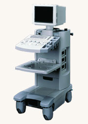
'Dedicated to ultrasound industry, Shantou Institute of Ultrasonic Instruments, Inc. (SIUI) has launched Apogee 3500, the Digital Color Doppler Ultrasound Imaging System. With latest imaging technologies, high-definition image quality and excellent practical functions, the Apogee 3500 offers optimal solutions for clinical ultrasonic examination.' 'The Apogee 3500 is available with many high-density, super broadband and multi-frequency probes, such as convex, micro-convex, linear, vaginal, rectal and phased array probes, which are widely applied for different clinical diagnoses, including abdomen (liver, kidney, gall-bladder, pancreas), gynecology (uterus, ovary), obstetrics (early pregnancy, basic OB, complete OB, multi gestation, fetal echo), cardiology (adult and pediatric cardiology), small parts (thyroid, galactophore, testicles, neonate), peripheral vascular and prostate.'
Device Information and Specification
CONFIGURATION
Normal system, color - gray scale(256)
Linear, convex and phased array
PROBES STANDARD
2.0 MHz ~ 12.0 MHz, broad band, tri-frequency
B-mode (B, 2B, 4B), M-mode, B/M-mode, real-time compound imaging, panoramic imaging, trapezoidal imaging (linear probes), spectrum Doppler (PWD and CWD), color Doppler flow imaging (CDFI), color power angio (CPA), tissue harmonic imaging (THI)
IMAGING OPTIONS
Real-time ZOOM, zoom rate and position selectable
OPTIONAL PACKAGE
H*W*D m
1.29 * 0.52 * 0.75
WEIGHT
110 kg
POWER REQUIREMENT
AC 220V/110V, 50Hz/60Hz
POWER CONSUMPTION
0.6 KVA
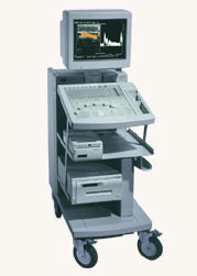
•  From Hitachi Medical Systems America Inc.;
The EUB-2000 B/W is a compact system capable of B-mode, M-mode, and optional with grayscale Doppler (EUB-2000 B/W Dop). This unit features intuitive operation, high mobility, and exceptional image quality. When color Doppler is not required, this system is an excellent choice for general purpose scanning. Device Information and Specification
APPLICATIONS
General purpose, OB/GYN, EUS, brachytherapy
CONFIGURATION
Compact system
Triple frequency
Linear, convex, radial, bi-plane, echoendoscope longitudinal
PROBE PORTS
Two
IMAGING OPTIONS
Simultaneous imaging with biplane probes
ADDITIONAL FEATURES
Cine / multi memory, application specific measuring, etc.
•  Hitachi Medical Systems America Inc.;
The EUB-525 is a mid-size ultrasound system with a full feature set. Capable of B-mode, M-mode, Doppler, color flow and color angiography it performs a variety of tasks in a simple and straightforward fashion. This system produces high quality images with excellent clarity and detail. Device Information and Specification
CLINICAL APPLICATION
CONFIGURATION
Compact, mobile system
Triple frequency
Linear, convex, radial, miniradial/miniprobe, bi-plane, echoendoscope longitudinal, echoendoscope radial
PROBE PORTS
Three
B/M-mode/simultaneous B/M-mode, CFM/CFA (color flow angiography)
IMAGING OPTIONS
Simultaneous imaging with biplane probes
IMAGING ENHANCEMENTS
Half-pitch scanning, dual scan line density, scan correlation and averaging, selectable receive filters
•
(TRUS) Transrectal sonography (also called transrectal ultrasonography, transrectal echography (TRE), endorectal ultrasound (ERUS or EUS)) is an ultrasound procedure used to examine the prostate gland, the rectum or bladder. A small, lubricated transducer placed into the rectum releases sound waves, which create echoes as they enter the region of interest. A computer creates a picture called a sonogram. TRUS is commonly used for guidance during a prostate needle biopsy and may be used to deliver brachytherapy and monitor cancer treatment. Transrectal ultrasonography detects enlargement, tumors and other abnormalities of the prostate, rectal polyps, rectal cancer, perianal infection, and sphincter muscle injuries. TRUS is also performed on male patients with infertility to view the prostate and surrounding structures and on patients with suspected bladder conditions or disease to view the bladder. See also Transurethral Sonography, Endoscopic Ultrasound, Pelvic Ultrasound, Rectal Probe, Biplane Probe, Endocavitary Echography and High Intensity Focused Ultrasound. Further Reading: News & More:
Result Pages : | Share This Page Look Ups |
Medical-Ultrasound-Imaging.com
former US-TIP.com
Member of SoftWays' Medical Imaging Group - MR-TIP • Radiology TIP • Medical-Ultrasound-Imaging
Copyright © 2008 - 2024 SoftWays. All rights reserved.
Terms of Use | Privacy Policy | Advertise With Us
former US-TIP.com
Member of SoftWays' Medical Imaging Group - MR-TIP • Radiology TIP • Medical-Ultrasound-Imaging
Copyright © 2008 - 2024 SoftWays. All rights reserved.
Terms of Use | Privacy Policy | Advertise With Us
[last update: 2023-11-06 01:42:00]