Medical Ultrasound Imaging
Tuesday, 14 May 2024
Info Sheets Out- side     | Ultrasound Database • 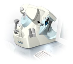 From Ultralink LLC;
'Artemis is a very high frequency (VHF) ultrasound eye scanner. In use, the patient leans forward placing their head onto an adjustable headrest. The headrest's unique design permits the patient to pull away quickly from the scanner if desired. An eyecup filled with a saline-based interface fluid couples the ultrasound signal to the eye, while a precision mechanism moves the transducer past the front of the eye. During the accurately controlled arc motion of the transducer, which lasts less than one second, many thousands of ultrasound samples are digitized. Following a scan, signal analysis is performed on a PC-compatible microcomputer, and the data are available for immediate viewing on an LCD monitor or disk storage. Artemis is very flexible; many adjustments to the scanning parameters are possible to customize the scan to your clinical needs. Functions are provided for centering the scan about the optical axis of the eye. The starting location of the scans as well as the extent can be varied as desired, to view image planes through the eye at different angles.' See also Ultrasound Biomicroscopy, A-Mode and A-Scan. Further Reading: News & More:
•
An image artifact is any image attribute, which is not present in the original imaged object. An image artifact is sometime the result of an improper operation of the imager, and in other times a consequence of natural processes or properties of the human body. Artifacts in diagnostic ultrasound are a reflection or an echo, which appears on the display and represents the real anatomical structure not correctly. An artifact can be a false, multiple or misleading information introduced by the imaging system or by interaction of ultrasound with the adjacent tissue. Artifacts in ultrasound can be classified as to their source like e.g.:
•
physiologic (motion, different sound velocities, acoustical impedances of tissue);
•
hardware (dimension of the ultrasound beam and the transducer array);
•
Image artifacts can occur in each medical ultrasound. Then an interpretation of the image is complicated and can eliminate the structural information of objects looking for. See also Ultrasound Imaging Procedures. •
Attenuation is the reduction of power, for example due to the passage through a medium or electrical component. In ultrasound imaging, attenuation means the decrease in amplitude and intensity as a sound wave travels through a medium. In ultrasound attenuation is often characterized as the half-value layer, or the half-power distance. These terms refer to the distance that ultrasound will travel in a particular tissue before its energy is attenuated to half its original value. Attenuation originates through:
•
divergence of the wavefront;
•
absorption of wave energy;
•
elastic reflection of wave energy;
•
elastic scattering of wave energy.
A thick muscled chest wall will offer a significant obstacle to the transmission of ultrasound. Non-muscle tissue such as fat does not attenuate acoustic energy as much. The half-value layer for bone is still less than muscle, that's why bone is such a barrier to ultrasound. See also Attenuation Coefficient, and Derated Quantity. Further Reading: Basics:
•
This coefficient is a quantification of the energy intensity loss of waves (electromagnetic or mechanical) due to attenuation.
In ultrasound imaging it is the relative energy intensity loss per traveled centimeter. The ultrasound attenuation coefficient is measured in units of dB/cm. The attenuation coefficient in soft tissues is nearly proportional to the ultrasound frequency. The attenuation coefficient is doubled when the frequency is doubled. This coefficient (dB/cm) divided by the frequency (MHz) is almost constant in a given tissue.
•
blood: 0.2 MHz x dB/cm;
•
fatty tissue: 0.6 MHz x dB/cm;
•
liver: 0.9 MHz x dB/cm;
•
soft tissue: 0.5-1.0 MHz x dB/cm.
•  From ESAOTE S.p.A.;
'The AU5 EPI is an ultrasound system for multi-disciplinary use that incorporates all the transducer technologies (linear, convex, phased array, and annular array) adapted to today's most diverse diagnostic requirements. Furthermore it offers the most modern and advanced techniques available today on the market.' | Share This Page Look Ups |
Medical-Ultrasound-Imaging.com
former US-TIP.com
Member of SoftWays' Medical Imaging Group - MR-TIP • Radiology TIP • Medical-Ultrasound-Imaging
Copyright © 2008 - 2024 SoftWays. All rights reserved.
Terms of Use | Privacy Policy | Advertise With Us
former US-TIP.com
Member of SoftWays' Medical Imaging Group - MR-TIP • Radiology TIP • Medical-Ultrasound-Imaging
Copyright © 2008 - 2024 SoftWays. All rights reserved.
Terms of Use | Privacy Policy | Advertise With Us
[last update: 2023-11-06 01:42:00]