Medical Ultrasound Imaging
Sunday, 19 May 2024
Info Sheets Out- side     | Ultrasound Database •
The definition of imaging is the visual representation of an object. Medical imaging is a broad term that encompasses various imaging modalities and techniques used in the field of medicine to visualize and study the body's anatomy and physiology. It includes both diagnostic and non-diagnostic imaging procedures, where diagnostic imaging specifically refers to the subset of medical imaging techniques that are primarily focused on diagnosing diseases or conditions. Medical imaging techniques are employed to obtain images or visual representations of the internal organs, tissues, and structures, aiding in the diagnosis, treatment, and monitoring of medical conditions.
The field of medical imaging has significantly evolved since the discovery of X-rays by Konrad Roentgen in 1896. Initially, radiological imaging involved focusing X-rays on the body and capturing the images on a single piece of film within a specialized cassette. Subsequent advancements introduced the use of fluorescent screens and special glasses for real-time visualization of X-ray images. A significant breakthrough came with the application of contrast agents, enhancing image contrast and improving organ visualization. In the 1950s, nuclear medicine studies utilizing gamma cameras demonstrated the uptake of low-level radioactive chemicals in organs, enabling the observation of biological processes in vivo. Currently, positron emission tomography (PET) and single photon emission computed tomography (SPECT) technologies play pivotal roles in clinical research and the diagnosis of biochemical and physiological processes. Additionally, the advent of the x-ray image intensifier in 1955 facilitated the capture and display of x-ray movies. In the 1960s, diagnostic imaging incorporated the principles of sonar, using ultrasonic waves generated by a quartz crystal. These waves, reflecting at the interfaces between different tissues, were received by ultrasound machines and translated into images through computer algorithms and reconstruction software. Ultrasound (ultrasonography) has become an indispensable diagnostic tool across various medical specialties, with immense potential for further advancements such as targeted contrast imaging, real-time 3D or 4D ultrasound, and molecular imaging. The first use of ultrasound contrast agents (USCA) dates back to 1968. Digital imaging techniques were introduced in the 1970s, revolutionizing conventional fluoroscopic image intensifiers. Godfrey Hounsfield's pioneering work led to the development of the first computed tomography (CT) scanner. Digital images are now electronic snapshots represented as grids of dots or pixels. X-ray CT brought about a breakthrough in medical imaging by providing cross-sectional images of the human body with high contrast between different types of soft tissue. These advancements were made possible by analog-to-digital converters and computers. The introduction of multislice spiral CT technology dramatically expanded the clinical applications of CT scans. The first magnetic resonance imaging (MRI) devices were tested on clinical patients in 1980. With technological improvements, such as higher field strength, more open MRI magnets, faster gradient systems, and novel data-acquisition techniques, MRI has emerged as a real-time interactive imaging modality capable of providing detailed structural and functional information of the body. Today, imaging in medicine offers a wide range of modalities, including:
•
X-ray projection imaging;
•
Fluoroscopy;
•
Computed tomography (CT / CAT);
•
Ultrasound imaging (US)
•
Magnetic resonance imaging (MRI), Magnetic source imaging (MSI);
•
Single photon emission computed tomography (SPECT);
•
Positron emission tomography (PET);
•
Mammography.
These imaging modalities have become integral components of modern healthcare. With the rapid advancement of digital imaging, efficient management has become important, leading to the expansion of radiology information systems (RIS) and the adoption of Picture Archiving and Communication Systems (PACS) for digital image archiving. In telemedicine, real-time transmission of all medical image modalities from MRI to X-ray, CT and ultrasound has become the standard. The field of medical imaging continues to evolve, promising further innovations and advancements in the future, ultimately contributing to improved patient care and diagnostics. See also History of Ultrasound Contrast Agents, and History of Ultrasound. • View NEWS results for 'Medical Imaging' (59). Further Reading: News & More:
•  'Founded in Seoul in 1985 by a team of research scientists from Korea's leading technology research institute, Medison rapidly established a reputation for innovation in digital imaging technology.
By 1994, the company had attained ISO 9001 certification for its ultrasound systems and had established a worldwide distribution network covering more than 80 countries'
'Founded in Seoul in 1985 by a team of research scientists from Korea's leading technology research institute, Medison rapidly established a reputation for innovation in digital imaging technology.
By 1994, the company had attained ISO 9001 certification for its ultrasound systems and had established a worldwide distribution network covering more than 80 countries'In 1998 Medison established a strategic alliance with Philips (formerly ATL, USA). Samsung Electronics acquired Medison in December 2010 on its way to become a top tier medical equipment manufacturer. In March 2011 the company got renamed to Samsung Medison. Ultrasound Systems: • 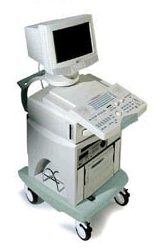 From ESAOTE S.p.A.;
'Megas CVX is your fully digital beamformer ultrasound system with phased, linear, convex and annular array technology. The modern modularity offers basic B-Mode, PW-CW Doppler, CFM and Power Doppler, TEI™-Tissue Enhancement Imaging and CnTI™- Contrast Tuned Imaging™.' • 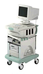 From ESAOTE S.p.A.;
'Megas GPX means all digital, all configurations, all probes feature a high standard level of quality and extended versatility. Based on digital beamformer technology, Megas GPX's functions can be easily updated to explore the new frontiers of the ultrasound world and give you the most flexible and powerful tool to make even the most difficult diagnoses.' •
(m) The SI base unit of distance. Definition: 1983 defined as the distance traveled by light in a vacuum during the time interval of 1/299 792 458 of a second. The speed of light in a vacuum, c, is one of the fundamental constants of nature. 1 meter (m) is equal to approximately 39.370 079 inches (in) 1 meter is equal to approximately 3.280 840 feet (ft) 1 meter is equal to approximately 1.093 613 3 yard (yd) 1 square meter (m2) is equal to approximately 10.763911 square feet (ft2) 1 inch = 2.54 centimeters Smaller or larger units are, e.g.: 1 (m) = 1 000 millimeter (mm) 1 kilometer (km) = 1 000 (m) 1 kilometer (km) = 0.62137 (statute) miles (mi) See also system international. • View NEWS results for 'Meter' (5). Further Reading: News & More:
| Share This Page Look Ups |
Medical-Ultrasound-Imaging.com
former US-TIP.com
Member of SoftWays' Medical Imaging Group - MR-TIP • Radiology TIP • Medical-Ultrasound-Imaging
Copyright © 2008 - 2024 SoftWays. All rights reserved.
Terms of Use | Privacy Policy | Advertise With Us
former US-TIP.com
Member of SoftWays' Medical Imaging Group - MR-TIP • Radiology TIP • Medical-Ultrasound-Imaging
Copyright © 2008 - 2024 SoftWays. All rights reserved.
Terms of Use | Privacy Policy | Advertise With Us
[last update: 2023-11-06 01:42:00]