Medical Ultrasound Imaging
Monday, 13 May 2024
Info Sheets Out- side     | 'Computer' p2 Searchterm 'Computer' found in 22 articles 22 definitions [ • ] Result Pages : •
Unlike regular sound, ultrasound can be directed into a single direction. The echoes received by a stationary probe will result in a single dimensional signal showing peaks for every major material change. To generate a 2D picture, the probe is swiveled, either mechanically or through a phased array of ultrasound transducers. The data is analyzed by computer and used to construct the image. In a similar way, 3D pictures can be generated by computer using a specialized probe. In this way, a photo of an unborn baby may be made. Some ultrasonography machines can produce color pictures, of sorts. Doppler ultrasonography is color coded onto a gray scale picture. From the amount of energy in each echo, the difference in acoustic impedance can be calculated and a color is then assigned accordingly. See also Densitometry and 3D Ultrasound. •
In 3D ultrasound (US) several 2D images are acquired by moving the probe across the body surface or rotating inserted probes. 3D-mode uses the same basic concept of a 2D ultrasound but rather than take the image from a single angle, the sonographer takes a volume image. The volume image that is displayed on the screen is a software rendering of all of the detected soft-tissue combined by specialized computer software to form three-dimensional images. The 3D volume rendering technique (VR) does not rely on segmentation (segmentation techniques are difficult to apply to ultrasound pictures) and makes it possible to obtain clear 3D ultrasound images for clinical diagnosis. A 3D ultrasound produces a still image. Diagnostic US systems with 3D display functions and linear array probes are mainly used for obstetric and abdominal applications. The combination of contrast agents, harmonic imaging and power Doppler greatly improves 3D US reconstructions. 3D imaging shows a better look at the organ being examined and is used for:
•
Detection of abnormal fetus development, e.g. of the face and limbs.
•
Visualization of e.g. the colon and rectum.
•
Pictures of blood flow in various organs or a fetus.
Fusion 3D imaging methods for generating compound images from two sets of ultrasound images (B-mode and Doppler images) enable the observation of the structural relationships between lesions and their associated blood vessels in three dimensions (maximum intensity projection). Further Reading: News & More:
•
(ADC) A system that receives analog input data and produces digital values at its output. Analog to digital converters are used by the ultrasound scanner to convert the received signal into a format more compatible with the computer systems. Ultrasound front-end-systems as well as many others sophisticated electronic systems use these analog signal processing components in connection with e.g., low-noise amplifier (LNA), and time gain compensation amplifier (TGC) as key elements in determining the overall system performance. See also Digital to Analog Converter, and Digitization. • 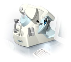 From Ultralink LLC;
'Artemis is a very high frequency (VHF) ultrasound eye scanner. In use, the patient leans forward placing their head onto an adjustable headrest. The headrest's unique design permits the patient to pull away quickly from the scanner if desired. An eyecup filled with a saline-based interface fluid couples the ultrasound signal to the eye, while a precision mechanism moves the transducer past the front of the eye. During the accurately controlled arc motion of the transducer, which lasts less than one second, many thousands of ultrasound samples are digitized. Following a scan, signal analysis is performed on a PC-compatible microcomputer, and the data are available for immediate viewing on an LCD monitor or disk storage. Artemis is very flexible; many adjustments to the scanning parameters are possible to customize the scan to your clinical needs. Functions are provided for centering the scan about the optical axis of the eye. The starting location of the scans as well as the extent can be varied as desired, to view image planes through the eye at different angles.' See also Ultrasound Biomicroscopy, A-Mode and A-Scan. Further Reading: News & More:
•  The company is a member of the Bracco Group, a highly innovative health care group and world leader in global integrated solutions for the diagnostic imaging field. The Bracco Group is headquartered in Milan, Italy. Its North American operations consist of Bracco Diagnostics and Bracco Research USA, both located in Princeton, New Jersey. Bracco Diagnostics is one of the fastest growing developers and marketers of diagnostic pharmaceuticals in North America, with products for various imaging applications, including Isovue® (iopamidol) X-ray contrast agent, ProHance®, (gadoteridol), MRI contrast agent, and nuclear medicine products.
Gadoteridol has been available in Europe and the USA for several years. Holder of the Marketing Authorization: Bracco International B.V. - Strawinskylaan 3051 - 1077 ZX Amsterdam The Netherlands. (Contact: Kirk Deeter, Phone: +NL-303-838-8708) The Bracco Group is the world's leading provider of global diagnostic imaging solutions, with net sales of more than 1 billion euro, of which more than 64% from international sales; it has operations in 115 countries and about 3,500 employees, of whom more than 600 work in R&D. Bracco invests around 15% of its turnover in R&D and has a portfolio of 1,500 patents worldwide. The Bracco Group deploys an integrated approach to diagnostic imaging, with an offer that encompasses contrast media, its core business where it is one of the world's top players, biomedical equipment from Esaote, one of the world's primary producers of magnetic resonance and ultrasonographic medical equipment, contrast media delivery systems from Acist Medical Systems, a top US company in advanced contrast media injection systems, and medical application software from EBIT-AET and Singapore's Volume Interactions, the leading developer of advanced medical software. Bracco has formed a high-level international research network, whose three centers in Milan, Geneva and Princeton study and develop products for the latest-generation diagnostic techniques, from X-rays and computerized axial tomography to magnetic resonance and echo contrast. Ultrasound Contrast Agents:
Contact Information
Please see Bracco Diagnostics, Inc.'s
Result Pages : | Share This Page Look Ups |
Medical-Ultrasound-Imaging.com
former US-TIP.com
Member of SoftWays' Medical Imaging Group - MR-TIP • Radiology TIP • Medical-Ultrasound-Imaging
Copyright © 2008 - 2024 SoftWays. All rights reserved.
Terms of Use | Privacy Policy | Advertise With Us
former US-TIP.com
Member of SoftWays' Medical Imaging Group - MR-TIP • Radiology TIP • Medical-Ultrasound-Imaging
Copyright © 2008 - 2024 SoftWays. All rights reserved.
Terms of Use | Privacy Policy | Advertise With Us
[last update: 2023-11-06 01:42:00]