Medical Ultrasound Imaging
Sunday, 19 May 2024
Info Sheets Out- side     | 'Display' p2 Searchterm 'Display' found in 81 articles 1 term [ • ] - 80 definitions [• ] Result Pages : •
B-scan comined with D-scan (D=Depth) is used to avoid image inhomogeneity. Different transmitter signals for each depth are applied and prefiltered pseudoinversely according to the transfer properties of the covering tissue. Pulse compression techniques with nonlinearly frequency modulated signals are used to gain the required energy for inverse filtering. D-scan is a modified C-scan used in nondestructive testing with the display of amplitudes. In the 2D graphical presentation, time of flight values are displayed in the top view on a test surface. See also A-Scan, B-Scan and C-Scan. • 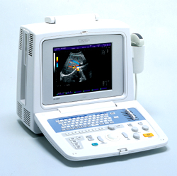 From Fukuda Denshi Co., Ltd.;
'Fully software controlled Image Processing (F-XT technology) Color Doppler & Power Doppler display available Autofocus function for transmission High-resolution SVGA display Cine memory with 128 frames Wide band Probe for multi frequency function 6-step STC adjustment DICOM-compatible Ethernet port 3.5-inch 640MB drive for image filing' •
Fetal ultrasound is a safe and non-invasive imaging technique used to visualize and monitor the development of a fetus during pregnancy. It employs high-frequency sound waves to create detailed images of the baby, the placenta, and the uterus. Fetal ultrasound provides valuable information about the baby's growth, organ development, and overall well-being. It is commonly used to determine gestational age, assess fetal anatomy, detect abnormalities, and monitor fetal movements and heart rate. This essential tool enables healthcare professionals to ensure the optimal health of both the mother and the baby throughout the pregnancy. The FDA (Food and Drug Administration) has established regulations governing ultrasound usage, including specific guidelines for fetal ultrasound examinations. These regulations permit an eight-fold increase in ultrasound intensity for fetal scans. They place considerably responsibility on the user to understand the output measurements, the mechanical index (MI), the thermal index (TI) and to use them in their scanning. The primary safety concern in prenatal diagnostic imaging is temperature rise. It is known that hyperthermia is teratogenic. The efforts of investigators have concentrated on defining the temperature increases and exposure times which may give rise to biological effects and on determining the ultrasound levels which might, in turn, lead to those temperature rises. In fetal ultrasound, the highest temperature increase would be expected to occur at bone and the thermal index with bone at/near the focus (TIB) would give the 'worst case' conditions. The mechanical index and thermal index must be displayed if the ultrasound system is capable of exceeding an index of 1. The displayed indices are based on the manufacturer's experimental and modeled data. However, an independent study has demonstrated significant discrepancies over declared spatial peak time averaged intensity (I-SPTA) output of up to 400%. See also ALARA Principle, Pregnancy Ultrasound and Doppler Fluximetry in Pregnancy. Further Reading: Basics:
•
Gray scale [also grayscale, grey scale = brit.] produces basically black and white images with series of shades of gray. Solid areas appear white and fluid areas appear black, varying from black at the weakest intensity to white at the strongest. Gray scale resolves artifacts as small as 1 mm. The display is made by transmitting bursts of energy and analyzing the returning signal. Gray scale pictures are limited to the gray scale tones; color pictures display more information because the color is added to the gray scale. Most ultrasound contrast agents also improve gray scale visualization of the flowing blood to such a degree that the tissue echogenicity increases. Gray scale enhancement of flow in an organ promises to improve lesion detection, along with the ability to differentiate between normal and abnormal areas, using many of the criteria already routinely used in CT and MRI. See also Compress, Densitometry, Triplex Exam and QB-Mode. •
(PACS) A system used to communicate and archive medical imaging data, mostly images and associated textural data generated in a radiology department, and disseminated throughout the hospital. A PACS is usually based on the DICOM (Digital Imaging and Communications in Medicine) standard. The main components in the PACS are: acquisition devices where the images are acquired;
•
short and longer term archives for storage of digital and textural data;
•
a database and database management;
•
diagnostic and review workstations;
•
software to run the system;
•
a communication network linking the system components;
•
interfaces with other networks (hospital and radiological information systems).
Acquisition devices, which acquire their data in direct digital format, like a MRI system, are most easily integrated into a PACS. Short term archives need to have rapid access, such as provided by a RAID (redundant array of independent disks), whereas long term archives need not have such rapid access and can be consigned, e.g. to optical disks or a magnetic. High speed networks are necessary for rapid transmission of imaging data from the short term archive to the diagnostic workstations. Optical fibre, ATM (asynchronous transfer mode), fast or switched Ethernet, are examples of high speed transmission networks, whereas demographic textural data may be transmitted along conventional Ethernet. Sophisticated software is a major element in any hospital-wide PACS. The software concepts include: preloading or prefetching of historical images pertinent to current examinations, worklists and folders to subdivide the vast mass of data acquired in a PACS in a form, which is easy and practical to access, default display protocols whereby images are automatically displayed on workstation monitors in a prearranged clinically logical order and format, and protocols radiologists can rapidly report worklists of undictated examinations, using a minimum of computer manipulation. Result Pages : | Share This Page Look Ups |
Medical-Ultrasound-Imaging.com
former US-TIP.com
Member of SoftWays' Medical Imaging Group - MR-TIP • Radiology TIP • Medical-Ultrasound-Imaging
Copyright © 2008 - 2024 SoftWays. All rights reserved.
Terms of Use | Privacy Policy | Advertise With Us
former US-TIP.com
Member of SoftWays' Medical Imaging Group - MR-TIP • Radiology TIP • Medical-Ultrasound-Imaging
Copyright © 2008 - 2024 SoftWays. All rights reserved.
Terms of Use | Privacy Policy | Advertise With Us
[last update: 2023-11-06 01:42:00]