Medical Ultrasound Imaging
Sunday, 19 May 2024
Info Sheets Out- side     | 'Gain' p3 Searchterm 'Gain' found in 19 articles 2 terms [ • ] - 17 definitions [• ] Result Pages : •
Phospholipids are a major component of all biological membranes. When placed in water, phospholipids form a bilayer, where the hydrophobic tails line up against each other. This forms a membrane with hydrophilic heads on both sides. This membrane is partially permeable and very flexible. Phospholipid containing microbubbles are in use as diagnostic ultrasound contrast agents. Phospholipids can be targeted to atheroma and other pathologic components to enhance atherosclerosis imaging. The majority of these echogenic liposomes range in diameter from 0.25 to 5.0 μm. •
Reject is a time gain compensation control that allows the sonographer to reduce the returning signal from more shallow structures in the body.
• 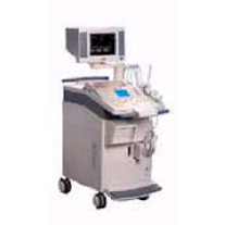 From Siemens Medical Systems;
'The SONOLINE Omnia™ ultrasound system offers mobility, high performance, and ease of use. This digital imaging system delivers excellent 2D, color flow, and Doppler image quality for a variety of general imaging exam types. In addition, the SONOLINE Omnia with cardiac option includes special features that enable use for adult cardiac imaging.'
Device Information and Specification
CLINICAL APPLICATION
Abdomen, small parts, pediatric, prostate, orthopedic, obstetrics, gynecology, cerebrovascular, musculoskeletal, rectal, peripheral vascular (venous and arterial), cardiology
CONFIGURATION
Compact, mobile system
Multi-Frequency and wideband
Wide range of linear/curved/phased array, mechanical, CW pencil probes, laparoscopic, intraoperative, biopsy, TEE transducers
PROBE PORTS
Five
IMAGING OPTIONS
OPTIONAL PACKAGE
Upgradeable applications, cardiac option
IMAGING ENHANCEMENTS
Ultra Fast 3D rendering
STORAGE
Magneto-Optical Drive of 640 MB
DATA PROCESSING
MultiDimensional image processor
• The field of medical imaging offers numerous career opportunities, and one profession is that of a sonographer. Sonographers play a critical role in healthcare by utilizing ultrasound technology to create images of the body's internal structures. •
Becoming a Sonographer: The educational and professional requirements for sonographers can vary from country to country. The duration of these programs can range from one to four years, depending on the country and level of qualification. The typical path in the United States begins with obtaining a post-secondary education in diagnostic medical sonography from an accredited program. These programs usually result in an associate's or bachelor's degree. Coursework typically covers anatomy, physiology, medical ethics, ultrasound physics, and specialized sonography techniques. Additionally, students gain practical experience through clinical internships in healthcare facilities. After completing their education, aspiring sonographers can choose to obtain professional certification through organizations such as the American Registry for Diagnostic Medical Sonography (ARDMS) or the American Registry of Radiologic Technologists (ARRT). Certification often requires passing examinations that assess knowledge and competency in specific areas of sonography. Many countries also have certification or registration requirements for sonographers. These certifications are typically obtained through professional bodies or organizations specific to each country. Examples include the Canadian Association of Registered Diagnostic Ultrasound Professionals (CARDUP) in Canada, the Australian Sonographers Accreditation Registry (ASAR) in Australia, and the Society and College of Radiographers (SCoR) in the United Kingdom. •
Job Description: Sonographers are skilled professionals who operate ultrasound machines and perform sonograms on patients. They work closely with physicians and other healthcare professionals to provide accurate and high-quality diagnostic images. Using sound waves, sonographers capture images of organs, tissues, and blood flow patterns, which are then used by medical practitioners to diagnose and monitor various medical conditions. Sonographers must have a comprehensive understanding of anatomy, physiology, and sonographic techniques to optimize image quality. They interact directly with patients, explaining procedures, addressing concerns, and ensuring patient comfort throughout the scanning process. Documentation of findings and communication with the medical team are also essential responsibilities. Some aspect of the job can be demanding, while sonographers often spend long hours on their feet, positioning and maneuvering patients during scans. Dealing with patients who are in pain, anxious, or difficult to scan requires empathy, patience, and excellent interpersonal skills. Sonographers often work in fast-paced environments, juggling multiple patients and procedures throughout the day. Effective time management is essential to ensure that scans are performed efficiently without compromising quality. Adhering to schedules and meeting the demands of the healthcare facility can add to the workload and stress levels. •
Salary Outlook: The salary of a sonographer can vary, based on factors such as experience, specialization, geographic location, and work setting. According to the U.S. Bureau of Labor Statistics, as of May 2021, the median annual wage for diagnostic medical sonographers was $77,740. Sonographers working in specialized hospitals, outpatient care centers, and diagnostic imaging centers tend to earn higher salaries compared to those in physician offices or government facilities. The salary prospects for sonographers outside the United States can vary significantly based on factors such as the country's economic conditions, healthcare system, demand for sonographers, and cost of living. •
Future Outlook: The future outlook for sonographers appears highly favorable. The demand for ultrasound imaging continues to grow due to advancements in medical technology and an aging population. This increasing demand for sonographers is expected to result in good job prospects and potential career advancement opportunities. Monitoring job markets, understanding regulatory requirements, and networking with professionals in international healthcare communities can provide valuable insights into future opportunities. See also Handheld Ultrasound, Ultrasound Machine, Sonography, Portable Ultrasound Machine, Ultrasound Accessories and Supplies, Environmental Protection and Ultrasound Technology. •
A flowmeter derives an accurate measure of the transit time ; transit time (or time of flight) is the difference in time for an ultrasonic pulse to travel with the direction of flow and against it. With bi-directional illumination it is the time taken for an ultrasound pulse to travel from one transducer to another. This time is used to measure blood flow rate. The difference between the upstream and downstream measured transit times calculates the volume flow. See also Echo Ranging. Result Pages : | Share This Page Look Ups |
Medical-Ultrasound-Imaging.com
former US-TIP.com
Member of SoftWays' Medical Imaging Group - MR-TIP • Radiology TIP • Medical-Ultrasound-Imaging
Copyright © 2008 - 2024 SoftWays. All rights reserved.
Terms of Use | Privacy Policy | Advertise With Us
former US-TIP.com
Member of SoftWays' Medical Imaging Group - MR-TIP • Radiology TIP • Medical-Ultrasound-Imaging
Copyright © 2008 - 2024 SoftWays. All rights reserved.
Terms of Use | Privacy Policy | Advertise With Us
[last update: 2023-11-06 01:42:00]