Medical Ultrasound Imaging
Sunday, 19 May 2024
Info Sheets Out- side     | 'Image Quality' p5 Searchterm 'Image Quality' found in 40 articles 1 term [ • ] - 39 definitions [• ] Result Pages : • 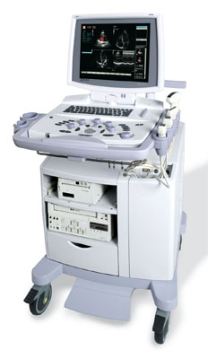 From Kontron Medical SAS;
Sigma 5000 Series 'THE ADVANCED VISION With the ability to continuously evolve over time for unprecedented return on investment, Imagic, Sigma 5000 Series, is the 2004 breakthrough in the ultrasound field. The all digital platform coupled with some proprietary technologies and a high quality, extra-large flat TFT monitor, display state-of-the-art image quality.' Specifications for this system will be available soon. •
An undesirable background interference or disturbance that affects image quality. The noise is commonly characterized by the standard deviation of signal intensity in the image of a uniform object (phantom) in the absence of artifacts. The measured noise may depend on the particular phantom used due to variable effects. Noisy images appear when the signal to noise ratio is too low. There are various noise sources in any electronic system, including Johnson noise, shot noise, thermal noise. See also Interference Artifact. •
In the field of medical ultrasound imaging, the term 'probe' specifically refers to the ultrasound transducer and represent the handheld device that emits and receives ultrasound waves during an examination. The probe encompasses various components such as the elements, backing material, electrodes, matching layer, and protective face that are responsible for both emitting and receiving the sound waves. Aperture, known also as the footprint, is the part of the probe that is in contact with the body. When the emitted sound waves encounter body tissues, they generate reflections that are received by the probe, which then generates a corresponding signal. In most cases, the probe emits ultrasound waves for only about 10% of the time and receives them for the remaining 90%. Probes are available in different shapes and sizes to accommodate various scanning situations. The footprint is linked to the arrangement of the piezoelectric crystals and comes in different shapes and sizes e.g. linear array transducer//convex transducer. The transducer plays a huge role in image quality and is one of the most expensive parts of the ultrasound machine. Mechanical probes steer the ultrasound beam driven by a motor and are capable of producing high-quality images, but they are prone to wear and tear. Mechanical probes have been mostly replaced by electronic multi-element transducers, but mechanical 3D probes still remain for abdominal and Ob-Gyn applications. In summary, the terms 'ultrasound transducer,' 'probe,' and 'scanhead' are often used interchangeably to refer to the same component of the ultrasound machine. Probes consist of multiple components and are available in different shapes and sizes depending on the sonographer's needs. See also Handheld Ultrasound, Ultrasound System Performance, Omnidirectional, Probe Cleaning, and Multi-frequency Probe, Further Reading: News & More:
•
Different sound velocities in tissue are causing refraction artifacts. With convex elastomer lens transducers, sound beam refraction at the skin interface can alter the transducer's focusing characteristics and beam profile, cause element to element nonuniformity, and cause phase changes in the acoustic wave. These cumulative refraction induced errors degrade the image quality through distortion and loss of resolution. Because the amount of refraction is proportional to the velocity mismatch, the greater the mismatch, the greater the refraction.
•  From Medison Co.,Ltd.;
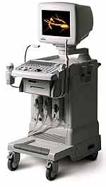
'It's the dawn of a new Evolution in affordable digital color ultrasound At Medison, we have a history of innovation. We were the first to make the leap to the third dimension when we introduced our groundbreaking digital 3D ultrasound platform with Live 3D™ technology in 1998, making it possible to capture true 3D volume data in real-time. Today, we're proud to introduce the SONOACE , the world's first true 3D color ultrasound system designed to bring the power of real-time 3D imaging to women's health specialists at a breakthrough price.' 'SONOACE 8000 Live PRIME, the true 3D ultrasound system designed to bring the power of live 3D in your hands. SONOACE 8000 Live PRIME offers superior image quality thanks to our new C-Square Technology and newly applied PSAD Beamformer.' Result Pages : | Share This Page Look Ups |
Medical-Ultrasound-Imaging.com
former US-TIP.com
Member of SoftWays' Medical Imaging Group - MR-TIP • Radiology TIP • Medical-Ultrasound-Imaging
Copyright © 2008 - 2024 SoftWays. All rights reserved.
Terms of Use | Privacy Policy | Advertise With Us
former US-TIP.com
Member of SoftWays' Medical Imaging Group - MR-TIP • Radiology TIP • Medical-Ultrasound-Imaging
Copyright © 2008 - 2024 SoftWays. All rights reserved.
Terms of Use | Privacy Policy | Advertise With Us
[last update: 2023-11-06 01:42:00]