Medical Ultrasound Imaging
Wednesday, 8 May 2024
'Pelvic Ultrasound' Searchterm 'Pelvic Ultrasound' found in 12 articles 1 term [ • ] - 10 definitions [• ] - 1 boolean [• ]Result Pages : • Pelvic Ultrasound
As far as ultrasound is concerned, 4D ultrasound (also referred to as live 3D ultrasound or 4B-mode) is the latest ultrasound technology - the fourth dimension means length, width, and depth over time. 4D Ultrasound takes 3D ultrasound images and adds the element of time to the progress so that a moving three-dimensional image is seen on the monitor. A 4D scan takes the same amounts of time as a 2D or 3D scan; the difference is the ultrasound equipment being used. One advantage of a 4D fetal ultrasound to a 2D-mode is that parents can see how their baby will generally look like. However, there are different opinions over the medical advantages. To scan a 3D ultrasound image, the probe is swept over the maternal abdomen. A computer takes multiple images and renders the 3D picture. With 4D imaging, the computer takes the images as multiple pictures while the probe is hold still and a 3D image is simultaneously rendered in real time on a monitor. In most cases, the standard 2D ultrasound is taken, and then the 3D/4D scan capability is added if an abnormality is detected or suspected. The 3D/4D sonogram is then focused on a specific area, to provide the details needed to assess and diagnose a suspected problem. A quick 4D scan of the face of the fetus may be performed at the end of a routine exam, providing the parents with a photo. See also Obstetric and Gynecologic Ultrasound, Pregnancy Ultrasound, Fetal Ultrasound and Abdominal Ultrasound. •
Gynecologic ultrasound and obstetric ultrasound are two distinct applications of ultrasound imaging that serve different purposes in the field of women's health. While both involve the use of ultrasound technology to examine the pelvic region, they have different focuses and objectives.
Gynecologic [gynaecologic, Brit.] ultrasound primarily concentrates on the evaluation of the female reproductive organs, including the uterus, ovaries, fallopian tubes, and surrounding structures. It is commonly performed for various gynecological concerns, such as abnormal bleeding, pelvic pain, infertility investigations, and monitoring of reproductive disorders. It can identify signs of inflammation, the presence of free fluid, cysts, and tumors. This non-invasive technique aids in diagnosing and monitoring gynecological pathologies, facilitating early intervention and appropriate treatment. Typically, a transabdominal sonogram is performed with a full bladder to provide an initial assessment. However, if the pelvic ultrasound reveals any abnormalities or fails to provide a clear image of the organs, a more detailed evaluation can be achieved through a transvaginal sonography. This approach allows for improved visualization of the uterus and ovaries by placing the ultrasound probe inside the vagina. Obstetric ultrasound, also known as prenatal, fetal or pregnancy ultrasound, is the branch of medical imaging that focuses on the use of ultrasound technology to assess the health and development of a fetus during pregnancy. Women with uncomplicated pregnancies commonly undergo an ultrasound examination between the 16th and 20th week of gestation. This routine assessment, performed with a real-time scanner, serves to determine accurate gestational age, monitor fetal size, and assess overall growth. The middle of the pregnancy trimester provides a crucial window for detecting many abnormalities of fetal anatomy. Advanced imaging techniques enable healthcare professionals to identify potential structural issues. Early detection of these abnormalities allows for timely intervention, counseling, and the implementation of appropriate management strategies. See also Pregnancy Ultrasound, Pelvic Ultrasound, Hysterosalpingo Contrast Sonography and Vaginal Probe. •
(AUS) Abdominal ultrasound, also known as abdominal sonography, is a medical imaging technique that focuses on the visualization and assessment of the abdominal organs. While 'abdominal ultrasound' is the commonly used term, there are alternative terms that can be used to refer to this imaging modality: (TAE) transabdominal echography, abdominal ultrasonography, sonogram, FAST (Focused Assessment with Sonography for Trauma). Abdominal ultrasound imaging is an invaluable clinical tool for identifying the underlying cause of abdominal pain. An abdominal ultrasound examination encompasses a comprehensive evaluation of the liver, gallbladder, biliary tree, pancreas, spleen, kidneys, and abdominal blood vessels. It is a cost-effective, safe, and non-invasive medical imaging modality that is typically utilized as the initial diagnostic investigation. Advanced ultrasound techniques, such as high-resolution ultrasound, endoscopic ultrasound, and contrast-enhanced Doppler, further enhance the detection of small lesions and provide detailed information for precise diagnosis. To prepare for an abdominal ultrasound, it is recommended to have nothing to eat or drink for at least 8 hours, starting from midnight the night before the examination. Indications:
•
Abdominal pain
•
Gallbladder or kidneys stones
•
Inflammation
•
Detection of cancer and metastasis
FAST (Focused Assessment with Sonography for Trauma) is a rapid diagnostic test used for trauma patients. It sequentially evaluates the presence of free fluid in the pericardium (hemopericardium) and in four specific views of the abdomen. These views include the right upper quadrant (RUQ), left upper quadrant (LUQ), subcostal, and suprapubic views. They aid in identifying hemoperitoneum in patients with potential truncal injuries. The space between the liver and the right kidney (RUQ), known as Morison's pouch, is a location where intraperitoneal fluid can accumulate. Emergency abdominal ultrasonography is indicated in cases of suspected aortic aneurysm, appendicitis, biliary and renal colic, as well as blunt or penetrating abdominal trauma. It plays a crucial role in the timely assessment and management of these conditions, providing critical information to guide appropriate treatment decisions. See also Handheld Ultrasound, Pelvic Ultrasound, Pregnancy Ultrasound, Prostate Ultrasound, Interventional Ultrasound and Pediatric Ultrasound. Further Reading: Basics: News & More:
•  From Verathon Inc.;
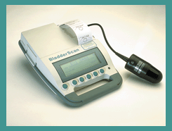
'The BladderScan® is a portable, noninvasive ultrasound instrument that measures bladder volume. It is battery-operated, easy to use, and quickly provides accurate results. In less than five minutes, you can use the BladderScan® to obtain a precise reading of a patient's bladder volume. The BladderScan® supplies the information that caregivers need to diagnose, treat and manage urinary conditions.' See also Urologic Ultrasound, Mirror Artifact, Pelvic Ultrasound, Transrectal Sonography and Ultrasonography. •  From Verathon Inc.;
'The BladderScan® BVI 6100 is a handheld, noninvasive ultrasound instrument that measures bladder volume. It is easy to operate, so any staff member can scan patients quickly and accurately. To view ultrasound images from exams and print BVI 6100 exam results for patient records and reimbursement, just log on to ScanPoint®, Verathon Inc.'s (formerly Diagnostic Ultrasound Corp.) innovative online service. The BVI 6100 makes it easier to provide the best care for your patients.' See also Urologic Ultrasound, Mirror Artifact, Pelvic Ultrasound, Transrectal Sonography and Ultrasonography. Result Pages : |
Medical-Ultrasound-Imaging.com
former US-TIP.com
Member of SoftWays' Medical Imaging Group - MR-TIP • Radiology TIP • Medical-Ultrasound-Imaging
Copyright © 2008 - 2024 SoftWays. All rights reserved.
Terms of Use | Privacy Policy | Advertise With Us
former US-TIP.com
Member of SoftWays' Medical Imaging Group - MR-TIP • Radiology TIP • Medical-Ultrasound-Imaging
Copyright © 2008 - 2024 SoftWays. All rights reserved.
Terms of Use | Privacy Policy | Advertise With Us
[last update: 2023-11-06 01:42:00]


