Medical Ultrasound Imaging
Sunday, 12 May 2024
Info Sheets Out- side     | 'Second' p3 Searchterm 'Second' found in 50 articles 1 term [ • ] - 49 definitions [• ] Result Pages : • 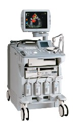 From ALOKA Co., Ltd.;
'A Platform for Pure Harmonic Detection Harmonic Echo™ technology constructs images using second harmonic components, which contain far less artifacts and noise than fundamental-frequency components. Pure Harmonic Detection is a technology to transmit distortion-free, fundamental-frequency ultrasound beams. And when the fundamental-frequency ultrasound beam is a pure sinusoidal wave, both the fundamental-frequency image and the Harmonic Echo™ image are much clearer with less noise. This technology is especially effective for obese patients and in a variety technically difficult scanning conditions.' •
The acoustic power of sound and ultrasound is the energy delivered per unit of time. The power is measured in Watt (W) and is proportional to the square of the amplitude. 1 W = 1 joule/second. See also Directivity Index, Spatial Average Intensity, and Source Level. •
(A or amp) The SI base unit of electric current. Definition: Two parallel conductors, infinitely long and having negligible cross section should be placed 1 meter apart in a perfect vacuum. One ampere is the current that creates between them a force of 0.2 microNewton per meter of length. One ampere represents a current flow of 1 coulomb of charge per second. One ampere of current results from a potential distribution of 1 volt per ohm of resistance, or from a power production rate of 1 watt per volt of potential. The unit is known informally as the amp, but A is its official symbol and is named for the French physicist André-Marie Ampère. See also System International. • 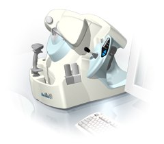 From Ultralink LLC;
'Artemis is a very high frequency (VHF) ultrasound eye scanner. In use, the patient leans forward placing their head onto an adjustable headrest. The headrest's unique design permits the patient to pull away quickly from the scanner if desired. An eyecup filled with a saline-based interface fluid couples the ultrasound signal to the eye, while a precision mechanism moves the transducer past the front of the eye. During the accurately controlled arc motion of the transducer, which lasts less than one second, many thousands of ultrasound samples are digitized. Following a scan, signal analysis is performed on a PC-compatible microcomputer, and the data are available for immediate viewing on an LCD monitor or disk storage. Artemis is very flexible; many adjustments to the scanning parameters are possible to customize the scan to your clinical needs. Functions are provided for centering the scan about the optical axis of the eye. The starting location of the scans as well as the extent can be varied as desired, to view image planes through the eye at different angles.' See also Ultrasound Biomicroscopy, A-Mode and A-Scan. Further Reading: News & More:
•  From Verathon Inc.;
'The BladderScan® BVM 6500 enables physicians quickly and noninvasively to determine UEBW (ultrasound estimated bladder weight) and bladder volume using 3-dimensional V-mode® ultrasound. The BVM 6500 was designed to assist in diagnosis of bladder hypertrophy secondary to obstruction.' See also Urologic Ultrasound, Mirror Artifact, Pelvic Ultrasound, Transrectal Sonography and Ultrasonography. Result Pages : | Share This Page Look Ups |
Medical-Ultrasound-Imaging.com
former US-TIP.com
Member of SoftWays' Medical Imaging Group - MR-TIP • Radiology TIP • Medical-Ultrasound-Imaging
Copyright © 2008 - 2024 SoftWays. All rights reserved.
Terms of Use | Privacy Policy | Advertise With Us
former US-TIP.com
Member of SoftWays' Medical Imaging Group - MR-TIP • Radiology TIP • Medical-Ultrasound-Imaging
Copyright © 2008 - 2024 SoftWays. All rights reserved.
Terms of Use | Privacy Policy | Advertise With Us
[last update: 2023-11-06 01:42:00]