Medical Ultrasound Imaging
Sunday, 19 May 2024
Info Sheets Out- side     | 'Sonography' p5 Searchterm 'Sonography' found in 61 articles 10 terms [ • ] - 51 definitions [• ] Result Pages : • 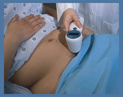 From Verathon Inc.;
'The BladderScan® BVM 6500 enables physicians quickly and noninvasively to determine UEBW (ultrasound estimated bladder weight) and bladder volume using 3-dimensional V-mode® ultrasound. The BVM 6500 was designed to assist in diagnosis of bladder hypertrophy secondary to obstruction.' See also Urologic Ultrasound, Mirror Artifact, Pelvic Ultrasound, Transrectal Sonography and Ultrasonography. •
Breast ultrasound (sonography or ultrasonography) it is an important tool in the characterization of breast lesions, detected with mammography or clinical breast examination. However, a breast sonogram is not approved by the U.S. Food and Drug Administration (FDA) as a screening tool for breast cancer and is used additional to a mammogram. Ultrasound is useful in guiding needles for fine needle aspiration and core biopsies. Breast ultrasound has optimal contrast resolution, but it lacks the spatial resolution of conventional mammography and cannot provide as much detail as a mammogram image. In addition, ultrasound is unable to show tiny calcium deposits (microcalcifications) that are often early indications of breast cancer. See also Biopsy, Interventional Ultrasound, Ultrasound Safety, Side Effect and Ultrasound Regulations. •
Contrast agents improve the sensitivity of vascular Doppler ultrasound, for example in cerebrovascular sonography or examinations of deep abdominal vessels. They also enlarge the role of transcranial Doppler. Microbubbles can be used with various modes e.g., color and power Doppler imaging, as well as pulsed-wave Doppler to increase the signal intensity. However, the ultrasound system must be suitable for contrast enhanced technology. Microbubbles usually stay within the vascular space; nevertheless, the contrast enhancement is limited to 2−6 minutes caused by physiologic clearance and bubble destruction. Depended on the application, contrast agents can be administered with a different injection rate e.g., bolus injection, slow injection, or continuous infusion. Stable, homogeneous, and prolonged enhancement can be obtained with perfusion, lasting until the infusion is stopped. See also Cerebrovascular Ultrasonography, Multiple Frame Trigger. •
From Bayer Schering Pharma AG: Echovist-200® was an effectively one-pass-only contrast medium for contrast sonography and Doppler-echocardiographic examinations for the detection, exclusion or follow-up of pathological states leading to hemodynamic changes. Because of the short intravascular life of the microparticles and microbubbles, transit through the pulmonary circulation is unusual. In cardiac evaluations Echovist-200® has been replaced by newer ultrasound contrast agents (USCA), therefore the manufacturing was discontinued. Another range of echo contrast application is the female genital tract, in particular for the demonstration or exclusion of acquired or congenital changes of the uterine cavity and for the visualization of the Fallopian tubes and investigation of their patency. 1 g Echovist-200 granules contain 1 g D-galactose microparticles. 1 ml aqueous solution for production of the suspension contains 200 mg D-galactose. Brand names in other countries: Ecovist.
Drug Information and Specification
RESEARCH NAME
-
DEVELOPER
INDICATION
Hysterosalpingo-contrast sonography (HyCoSy), echocardiographic use in neonates and children
APPLICATION
Intravenous injection
TYPE
Microbubble
D-GALACTOSE®
Air
MICROBUBBLE SIZE
99 % < 12 μm, 95 % < 8 μm
STORAGE
Store below 30 °C
PRESENTATION
Vials of 20 ml with 3.0 g granulate incl. one vial of 15 ml containing 13.5 ml D-galactose solution, one mini-spike
PREPARATION
Reconstitute with water
DO NOT RELY ON THE INFORMATION PROVIDED HERE, THEY ARE
NOT A SUBSTITUTE FOR THE ACCOMPANYING PACKAGE INSERT! Further Reading: News & More:
•
Ultrasonography of the kidneys (renal ultrasound) is part of a complete examination of the abdomen. Ultrasound is used to determine the size, shape, and exact position of the kidneys. Renal ultrasound provides important information regarding kidney function, related blood vessels, kidney stones, renal cysts, tumors, or hydronephrosis (suggestive of obstruction or blockage of the kidney). The kidneys are scanned on longitudinal and transverse planes. Patients should avoid carbonated drinks such as soda or seltzer the day before, and have a full bladder for the test. Lithotripsy is a therapeutic ultrasound procedure used to shatter simple stones in the kidney or upper urinary tract. See also Urologic Ultrasound, Reflux Sonography. Result Pages : | Share This Page Look Ups |
Medical-Ultrasound-Imaging.com
former US-TIP.com
Member of SoftWays' Medical Imaging Group - MR-TIP • Radiology TIP • Medical-Ultrasound-Imaging
Copyright © 2008 - 2024 SoftWays. All rights reserved.
Terms of Use | Privacy Policy | Advertise With Us
former US-TIP.com
Member of SoftWays' Medical Imaging Group - MR-TIP • Radiology TIP • Medical-Ultrasound-Imaging
Copyright © 2008 - 2024 SoftWays. All rights reserved.
Terms of Use | Privacy Policy | Advertise With Us
[last update: 2023-11-06 01:42:00]