Medical Ultrasound Imaging
Thursday, 9 May 2024
Info Sheets Out- side     | 'Spatial Resolution' p2 Searchterm 'Spatial Resolution' found in 15 articles 1 term [ • ] - 9 definitions [• ] - 5 booleans [• ]Result Pages : •
(CHI) Contrast harmonic imaging is an ultrasound technique to improve the measurement of blood perfusion or capillary blood flow. Based on the nonlinear properties of contrast agents, CHI transmits at the fundamental frequency but receives at the second harmonic. Contrast enhanced echo signals contain significant energy components at higher harmonics (bubbles acts as harmonic oscillators), while tissue echoes do not. Caused by that contrast signal can be separated from tissue echoes by the characteristic signal. In combination with the pulse inversion technique, CHI promises very high contrast agent sensitivity with high spatial resolution. See also Ultrasound Contrast Agent Safety and Hemoglobin. Further Reading: Basics:
•
QB-mode (Quadratic Brightness-mode) images are gray scale images from the quadratic component. QB-mode achieves higher contrast and increased dynamic range than the standard B-mode ultrasound images, without loss in spatial resolution.
Further Reading: Basics:
• 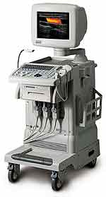 From Medison Co.,Ltd.;
'SONOACE 8000 EX PRIME, combines evolutionary C-Square technology and PSAD Beamformer in addition to our next-generation digital beamforming technology with state-of-the-art intelligent digital signal processing. SONOACE 8000 EX PRIME creates images with outstanding detail and spatial resolution that will add new clarity to your diagnoses.' •
Ultrasound biomicroscopy utilizes high frequency (10 - 50 MHz) diagnostic ultrasound to examine living tissue at a microscopic level and allows to image the skin with extremely high resolution to a depth of 2-3 centimeters. Ultrasound biomicroscopy images provide detailed anatomical information that can lead to better and more accurate treatments and avoid a biopsy. Ultrasound biomicroscopy improves also the spatial resolution of US images of the anterior segment of the eye. US biomicroscopy of the eye operates in the 50 MHz range with a possible axial resolution on the order of 30 μm. In this frequency range, tissue penetration of only approximately 5 mm is attainable. Both continuous wave Doppler and high-frequency pulsed Doppler can be used. See also Ultrasound Imaging Procedures, A-Scan, B-Scan and C-Scan. Further Reading: News & More:
•
A phantom is used to control the imaging performance of ultrasound transducers. The spatial resolution, dead zone, linear fidelity, depth of penetration and image uniformity is important for the image quality. For the axial and lateral resolution, the standard definition is the resolution of objects parallel and perpendicular to the path of the sound beam. Ultrasound pictures created by scans of specially designed ultrasound phantoms can quantify the imaging quality and transducer performance. Phantoms contain one or more materials that simulate a tissue in its interaction with ultrasound. Several phantoms are available specifically for quality control. Transducer characterization consists of a standard pulse echo analysis and insertion loss measurement for each probe. The quality variation from the baseline should be tracked over a period. Further Reading: Basics: Result Pages : | Share This Page Look Ups |
Medical-Ultrasound-Imaging.com
former US-TIP.com
Member of SoftWays' Medical Imaging Group - MR-TIP • Radiology TIP • Medical-Ultrasound-Imaging
Copyright © 2008 - 2024 SoftWays. All rights reserved.
Terms of Use | Privacy Policy | Advertise With Us
former US-TIP.com
Member of SoftWays' Medical Imaging Group - MR-TIP • Radiology TIP • Medical-Ultrasound-Imaging
Copyright © 2008 - 2024 SoftWays. All rights reserved.
Terms of Use | Privacy Policy | Advertise With Us
[last update: 2023-11-06 01:42:00]