Medical Ultrasound Imaging
Wednesday, 8 May 2024
Info Sheets Out- side     | '3D Ultrasound' p5 Searchterm '3D Ultrasound' found in 23 articles 1 term [ • ] - 18 definitions [• ] - 4 booleans [• ]Result Pages : • 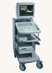 Hitachi Medical Systems America Inc.;
The EUB-525 is a mid-size ultrasound system with a full feature set. Capable of B-mode, M-mode, Doppler, color flow and color angiography it performs a variety of tasks in a simple and straightforward fashion. This system produces high quality images with excellent clarity and detail. Device Information and Specification
CLINICAL APPLICATION
CONFIGURATION
Compact, mobile system
Triple frequency
Linear, convex, radial, miniradial/miniprobe, bi-plane, echoendoscope longitudinal, echoendoscope radial
PROBE PORTS
Three
B/M-mode/simultaneous B/M-mode, CFM/CFA (color flow angiography)
IMAGING OPTIONS
Simultaneous imaging with biplane probes
IMAGING ENHANCEMENTS
Half-pitch scanning, dual scan line density, scan correlation and averaging, selectable receive filters
•
In the field of medical ultrasound imaging, the term 'probe' specifically refers to the ultrasound transducer and represent the handheld device that emits and receives ultrasound waves during an examination. The probe encompasses various components such as the elements, backing material, electrodes, matching layer, and protective face that are responsible for both emitting and receiving the sound waves. Aperture, known also as the footprint, is the part of the probe that is in contact with the body. When the emitted sound waves encounter body tissues, they generate reflections that are received by the probe, which then generates a corresponding signal. In most cases, the probe emits ultrasound waves for only about 10% of the time and receives them for the remaining 90%. Probes are available in different shapes and sizes to accommodate various scanning situations. The footprint is linked to the arrangement of the piezoelectric crystals and comes in different shapes and sizes e.g. linear array transducer//convex transducer. The transducer plays a huge role in image quality and is one of the most expensive parts of the ultrasound machine. Mechanical probes steer the ultrasound beam driven by a motor and are capable of producing high-quality images, but they are prone to wear and tear. Mechanical probes have been mostly replaced by electronic multi-element transducers, but mechanical 3D probes still remain for abdominal and Ob-Gyn applications. In summary, the terms 'ultrasound transducer,' 'probe,' and 'scanhead' are often used interchangeably to refer to the same component of the ultrasound machine. Probes consist of multiple components and are available in different shapes and sizes depending on the sonographer's needs. See also Handheld Ultrasound, Ultrasound System Performance, Omnidirectional, Probe Cleaning, and Multi-frequency Probe, Further Reading: News & More:
•
Ultrasound images can be manipulated for evaluation in various ways. Post processing includes: 3D imaging analysis, multi planar reconstruction (MPR), maximum intensity projection (MIP), etc.
Further Reading: News & More:
Result Pages : | Share This Page Look Ups |
Medical-Ultrasound-Imaging.com
former US-TIP.com
Member of SoftWays' Medical Imaging Group - MR-TIP • Radiology TIP • Medical-Ultrasound-Imaging
Copyright © 2008 - 2024 SoftWays. All rights reserved.
Terms of Use | Privacy Policy | Advertise With Us
former US-TIP.com
Member of SoftWays' Medical Imaging Group - MR-TIP • Radiology TIP • Medical-Ultrasound-Imaging
Copyright © 2008 - 2024 SoftWays. All rights reserved.
Terms of Use | Privacy Policy | Advertise With Us
[last update: 2023-11-06 01:42:00]