Medical Ultrasound Imaging
Sunday, 19 May 2024
Info Sheets Out- side     | 'Ultrasonic' p5 Searchterm 'Ultrasonic' found in 40 articles 4 terms [ • ] - 36 definitions [• ] Result Pages : •
The dwell time (also called ensemble length or packet size) is the transmitting duration of ultrasonic waves focused at one place or the Doppler samples for each line of sight. The reduction of the dwell time is of use if threshold pressures are exceeded. The sensitivity to slow flow and accuracy of Doppler measurement increase with longer dwell times. With increased dwell times, the frame rates decrease and the capability of Color Doppler images to depict fast changes in hemodynamics is limited. • 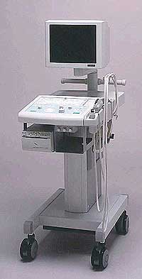 From Fukuda Denshi Co., Ltd.;
'Operation-friendly Switches and Controls backlit to permit the operator to identify them in a darkroom Image Optimization through Ultrasonic Frequency Changeover without replacement of the probe High frame rate enhancement mode for Cardiac echo Cine memory for selection of best data through SCROLL, LOOP, or MANUAL review Programmable function keys allow to get predefined for measurement parameters. Special measurement software usable in a wide range. Measurement Report page on the display can be printed as required.' •
(MI) The mechanical index is an estimate of the maximum amplitude of the pressure pulse in tissue. It is an indicator of the likelihood of mechanical bioeffects (streaming and cavitation). The mechanical index of the ultrasound beam is the amount of negative acoustic pressure within a ultrasonic field and is used to modulate the output signature of US contrast agents and to incite different microbubble responses. The mechanical index is defined as the peak rarefactional pressure (negative pressure) divided by the square root of the ultrasound frequency. The FDA ultrasound regulations allow a mechanical index of up to 1.9 to be used for all applications except ophthalmic (maximum 0.23). The used range varies from 0.05 to 1.9. At low acoustic power, the acoustic response is considered as linear. At a low MI (less than 0.2), the microbubbles undergo oscillation with compression and rarefaction that are equal in amplitude and no special contrast enhanced signal is created. Microbubbles act as strong scattering objects due to the difference in impedance between air and liquid, and the acoustic response is optimized at the resonant frequency of a microbubble. At higher acoustic power (MI between 0.2-0.5), nonlinear oscillation occurs preferentially with the bubbles undergoing rarefaction that is greater than compression. Ultrasound waves are created at harmonics of the delivered frequency. The harmonic response frequencies are different from that of the incident wave (fundamental frequency) with subharmonics (half of the fundamental frequency), harmonics (including the second harmonic response at twice the fundamental frequency), and ultra-harmonics obtained at 1.5 or 2.5 times the fundamental frequency. These contrast enhanced ultrasound signals are microbubble-specific. At high acoustic power (MI greater than 0.5), microbubble destruction begins with emission of high intensity transient signals very rich in nonlinear components. Intermittent imaging becomes needed to allow the capillaries to be refilled with fresh microbubbles. Microbubble destruction occurs to some degree at all mechanical indices. A mechanical index from 0.8 to 1.9 creates high microbubble destruction. The output signal is unique to the contrast agent. Further Reading: Basics:
News & More:
•
The definition of imaging is the visual representation of an object. Medical imaging is a broad term that encompasses various imaging modalities and techniques used in the field of medicine to visualize and study the body's anatomy and physiology. It includes both diagnostic and non-diagnostic imaging procedures, where diagnostic imaging specifically refers to the subset of medical imaging techniques that are primarily focused on diagnosing diseases or conditions. Medical imaging techniques are employed to obtain images or visual representations of the internal organs, tissues, and structures, aiding in the diagnosis, treatment, and monitoring of medical conditions.
The field of medical imaging has significantly evolved since the discovery of X-rays by Konrad Roentgen in 1896. Initially, radiological imaging involved focusing X-rays on the body and capturing the images on a single piece of film within a specialized cassette. Subsequent advancements introduced the use of fluorescent screens and special glasses for real-time visualization of X-ray images. A significant breakthrough came with the application of contrast agents, enhancing image contrast and improving organ visualization. In the 1950s, nuclear medicine studies utilizing gamma cameras demonstrated the uptake of low-level radioactive chemicals in organs, enabling the observation of biological processes in vivo. Currently, positron emission tomography (PET) and single photon emission computed tomography (SPECT) technologies play pivotal roles in clinical research and the diagnosis of biochemical and physiological processes. Additionally, the advent of the x-ray image intensifier in 1955 facilitated the capture and display of x-ray movies. In the 1960s, diagnostic imaging incorporated the principles of sonar, using ultrasonic waves generated by a quartz crystal. These waves, reflecting at the interfaces between different tissues, were received by ultrasound machines and translated into images through computer algorithms and reconstruction software. Ultrasound (ultrasonography) has become an indispensable diagnostic tool across various medical specialties, with immense potential for further advancements such as targeted contrast imaging, real-time 3D or 4D ultrasound, and molecular imaging. The first use of ultrasound contrast agents (USCA) dates back to 1968. Digital imaging techniques were introduced in the 1970s, revolutionizing conventional fluoroscopic image intensifiers. Godfrey Hounsfield's pioneering work led to the development of the first computed tomography (CT) scanner. Digital images are now electronic snapshots represented as grids of dots or pixels. X-ray CT brought about a breakthrough in medical imaging by providing cross-sectional images of the human body with high contrast between different types of soft tissue. These advancements were made possible by analog-to-digital converters and computers. The introduction of multislice spiral CT technology dramatically expanded the clinical applications of CT scans. The first magnetic resonance imaging (MRI) devices were tested on clinical patients in 1980. With technological improvements, such as higher field strength, more open MRI magnets, faster gradient systems, and novel data-acquisition techniques, MRI has emerged as a real-time interactive imaging modality capable of providing detailed structural and functional information of the body. Today, imaging in medicine offers a wide range of modalities, including:
•
X-ray projection imaging;
•
Fluoroscopy;
•
Computed tomography (CT / CAT);
•
Ultrasound imaging (US)
•
Magnetic resonance imaging (MRI), Magnetic source imaging (MSI);
•
Single photon emission computed tomography (SPECT);
•
Positron emission tomography (PET);
•
Mammography.
These imaging modalities have become integral components of modern healthcare. With the rapid advancement of digital imaging, efficient management has become important, leading to the expansion of radiology information systems (RIS) and the adoption of Picture Archiving and Communication Systems (PACS) for digital image archiving. In telemedicine, real-time transmission of all medical image modalities from MRI to X-ray, CT and ultrasound has become the standard. The field of medical imaging continues to evolve, promising further innovations and advancements in the future, ultimately contributing to improved patient care and diagnostics. See also History of Ultrasound Contrast Agents, and History of Ultrasound. Further Reading: News & More:
•
Microbubbles filled with air or inert gases are used as contrast agents in ultrasound imaging. Compression and rarefaction created by an ultrasound wave insonating a gas-filled microbubble along with the mechanical index of the ultrasonic beam lead to volume pulsations of the bubbles, and it is this change that results in the signal enhancement. Microbubbles have diameters from 1 μm to 10 μm and a thin flexible or rigid shell composed of albumin, lipid, or polymer confining a gas such as nitrogen, or a perfluorocarbon. These microbubbles can cross the pulmonary capillaries and have a serum half-life of a few minutes. Microbubbles in the 1-10 μm range have their resonance at the frequencies used in diagnostic ultrasound (1−15MHz). Smaller bubbles resonate at higher frequencies. Caused by this coincidence, they are such effective reflectors. The intrinsic compressibility of microbubbles is approximately 17,000 times more than water, and they are very strong scatterers of ultrasound. Under acoustic pressure the vibrating bubble radius may have a conventional linear response or a harmonic non-linear response. Microbubbles usually increase the Doppler signal amplitude by up to 30 dB. Further Reading: Basics:
News & More:
Result Pages : | Share This Page Look Ups |
Medical-Ultrasound-Imaging.com
former US-TIP.com
Member of SoftWays' Medical Imaging Group - MR-TIP • Radiology TIP • Medical-Ultrasound-Imaging
Copyright © 2008 - 2024 SoftWays. All rights reserved.
Terms of Use | Privacy Policy | Advertise With Us
former US-TIP.com
Member of SoftWays' Medical Imaging Group - MR-TIP • Radiology TIP • Medical-Ultrasound-Imaging
Copyright © 2008 - 2024 SoftWays. All rights reserved.
Terms of Use | Privacy Policy | Advertise With Us
[last update: 2023-11-06 01:42:00]