Medical Ultrasound Imaging
Sunday, 19 May 2024
Info Sheets Out- side     | 'Ultrasound Imaging Modes' p5 Searchterm 'Ultrasound Imaging Modes' found in 23 articles 1 term [ • ] - 5 definitions [• ] - 17 booleans [• ]Result Pages : • 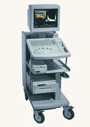 Hitachi Medical Systems America Inc.;
The EUB-525 is a mid-size ultrasound system with a full feature set. Capable of B-mode, M-mode, Doppler, color flow and color angiography it performs a variety of tasks in a simple and straightforward fashion. This system produces high quality images with excellent clarity and detail. Device Information and Specification
CLINICAL APPLICATION
CONFIGURATION
Compact, mobile system
Triple frequency
Linear, convex, radial, miniradial/miniprobe, bi-plane, echoendoscope longitudinal, echoendoscope radial
PROBE PORTS
Three
B/M-mode/simultaneous B/M-mode, CFM/CFA (color flow angiography)
IMAGING OPTIONS
Simultaneous imaging with biplane probes
IMAGING ENHANCEMENTS
Half-pitch scanning, dual scan line density, scan correlation and averaging, selectable receive filters
•
(CFI) Color flow imaging is based on pulsed ultrasound Doppler technology. With this technique multiple sample volumes among multiple planes are detected and a color map for direction and velocity flow data is displayed. Common mapping formats are BART (Blue Away, Red Towards) or RABT (Red Away, Blue Towards). Enhanced or variance flow maps show saturations and intensities that indicate higher velocities and turbulence or acceleration. Some maps utilize a third color (green) to indicate accelerating velocities and turbulence. Color flow Doppler imaging is not as precise as conventional Doppler and is best used to scan a larger area and then use other Doppler modes to obtain more precise data. See also Color Amplitude Imaging, Color Priority, and Color Saturation. •
A-mode (Amplitude-mode) ultrasound is a technique used to assess organ dimensions and determine the depth of an organ. While A-mode technology was previously employed in midline echoencephalography for rapid screening of intracranial mass lesions and ophthalmologic scanning, it is now considered obsolete in medical imaging. Nonetheless, the A-mode scan has found applications in early pregnancy assessment (specifically the detection of fetal heartbeats), cephalometry, and placental localization.
When the ultrasound beam encounters an anatomic boundary, the received sound impulse is processed to appear as a vertical reflection of a point. On the display, it looks like spikes of different heights (the amplitude). The intensity of the returning impulse determined the height of the vertical reflection and the time it took for the impulse to make the round trip would determine the space between verticals. The distance between these spikes can be measured accurately by dividing the speed of sound in tissue (1540 m/sec) by half the sound travel time. During an echoencephalography scan, the first A-mode scan is acquired from the right side of the head and captured on film. Subsequently, the probe is positioned at the corresponding point on the left side, and a second exposure is captured on the same film, displaying inverted spikes. The A-mode ultrasound could be used to identify structures normally located in the midline of the brain such as the third ventricle and falx cerebri. The midline structures would be aligned in normal patients but show displacement in patients with mass lesion such as a subdural, epidural, or intracranial hemorrhage. See also 2D Ultrasound, 3D Ultrasound, 4D Ultrasound, Ultrasound Biomicroscopy, A-scan, B-mode and the Infosheet about ultrasound modes. Result Pages : | Share This Page Look Ups |
Medical-Ultrasound-Imaging.com
former US-TIP.com
Member of SoftWays' Medical Imaging Group - MR-TIP • Radiology TIP • Medical-Ultrasound-Imaging
Copyright © 2008 - 2024 SoftWays. All rights reserved.
Terms of Use | Privacy Policy | Advertise With Us
former US-TIP.com
Member of SoftWays' Medical Imaging Group - MR-TIP • Radiology TIP • Medical-Ultrasound-Imaging
Copyright © 2008 - 2024 SoftWays. All rights reserved.
Terms of Use | Privacy Policy | Advertise With Us
[last update: 2023-11-06 01:42:00]