Medical Ultrasound Imaging
Sunday, 19 May 2024
Info Sheets Out- side     | 'Ultrasound imaging' p11 Searchterm 'Ultrasound imaging' found in 68 articles 3 terms [ • ] - 65 definitions [• ] Result Pages : •
In the field of medical ultrasound imaging, the term 'probe' specifically refers to the ultrasound transducer and represent the handheld device that emits and receives ultrasound waves during an examination. The probe encompasses various components such as the elements, backing material, electrodes, matching layer, and protective face that are responsible for both emitting and receiving the sound waves. Aperture, known also as the footprint, is the part of the probe that is in contact with the body. When the emitted sound waves encounter body tissues, they generate reflections that are received by the probe, which then generates a corresponding signal. In most cases, the probe emits ultrasound waves for only about 10% of the time and receives them for the remaining 90%. Probes are available in different shapes and sizes to accommodate various scanning situations. The footprint is linked to the arrangement of the piezoelectric crystals and comes in different shapes and sizes e.g. linear array transducer//convex transducer. The transducer plays a huge role in image quality and is one of the most expensive parts of the ultrasound machine. Mechanical probes steer the ultrasound beam driven by a motor and are capable of producing high-quality images, but they are prone to wear and tear. Mechanical probes have been mostly replaced by electronic multi-element transducers, but mechanical 3D probes still remain for abdominal and Ob-Gyn applications. In summary, the terms 'ultrasound transducer,' 'probe,' and 'scanhead' are often used interchangeably to refer to the same component of the ultrasound machine. Probes consist of multiple components and are available in different shapes and sizes depending on the sonographer's needs. See also Handheld Ultrasound, Ultrasound System Performance, Omnidirectional, Probe Cleaning, and Multi-frequency Probe, Further Reading: News & More:
•
The elements of a rectangular array transducer (also called matrix transducer) are arranged in a rectangular pattern. Rectangular arrays with unequal rows (e.g. 3, 5, 7) of transducer elements are in real 2D (two-dimensional), but they are termed 1.5D, because the number of rows is much less than the number of columns. Their main advantage is electronic focusing even in the elevation plane (z-plane). The transducers that are termed 2D have an equal number of rows and columns. 2D transducers have the potential to provide real-time 3D ultrasound imaging without moving the transducer. Active matrix array transducers have several elements in the short axis and in addition multiple elements along the long axis. This allows electronic focusing in both axes, resulting in a narrower elevation axis beam width in the near field and far field. •
Reflection of the sound beam occurs when it hits a boundary between materials having different acoustic impedance. The reflection (echo) is the portion of a sound that is returned from the boundary. The reflection time (the time taken for the wave to return to the probe) can be used to determine the depth of the object.
The reflection within the body produces the ultrasound image, but should be minimized at an ultrasound couplant to skin boundary where the couplant acts as an acoustic window through which the image is seen. The amount of sound waves, which are reflected back at the interface between two tissues is depend on the angle of incidence and the difference between the acoustic impedance values of the two tissues. If the difference is great, a large part of the sound waves will be reflected back. If too much sound is reflected back and not enough waves are remaining to be able to penetrate the tissue, the imaging will be poor. If the difference is small, a small amount will be reflected back. Enough sound signal remains to continue with ultrasound imaging. If the ultrasound beam meets a rough surface or small object, the beam is scattered in all directions and only a small amount will be received by the probe. See also False Distance Artifact, Target Strength, and Snells Law. •
Reflux sonography, as an alternative to micturating cystography (MCU), evaluates vesico-ureteral reflux (VUR), a common problem in children. Contrast enhanced pulse-inversion imaging shows best results. During the instillation of an ultrasound contrast agent into the bladder, (as for a conventional MCU) the lower ureters and renal pelves are scanned transabdominally as the bladder is filled to stimulate micturition. Advantages for reflux sonography are a high sensitivity and the avoidance of X-rays. A disadvantage is the poorer depiction of the posterior urethra. However, for girls and for all follow-up studies, the ultrasound MCU has become standard in many pediatric ultrasound departments. See also Urologic Ultrasound, Kidney Ultrasound, Ultrasound Safety, Ultrasound Imaging Modes. • 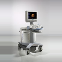 From Siemens Medical Systems;
'The Antares™ system was built on four foundations that mark the beginning of a new paradigm in ultrasound imaging: Diagnostic Precision, DIMAQ-IP, ErgoDynamic™ imaging system design, and Workflow Control.' Specifications for this system will be available soon. Result Pages : | Share This Page Look Ups |
Medical-Ultrasound-Imaging.com
former US-TIP.com
Member of SoftWays' Medical Imaging Group - MR-TIP • Radiology TIP • Medical-Ultrasound-Imaging
Copyright © 2008 - 2024 SoftWays. All rights reserved.
Terms of Use | Privacy Policy | Advertise With Us
former US-TIP.com
Member of SoftWays' Medical Imaging Group - MR-TIP • Radiology TIP • Medical-Ultrasound-Imaging
Copyright © 2008 - 2024 SoftWays. All rights reserved.
Terms of Use | Privacy Policy | Advertise With Us
[last update: 2023-11-06 01:42:00]