Medical Ultrasound Imaging
Sunday, 20 September 2026

 Info Sheets  Out- side     | 'Mode' p12 Searchterm 'Mode' found in 134 articles 14 terms [ • ] - 120 definitions [• ] Result Pages : •
(CD) Color Doppler is an ultrasound imaging mode, which visualizes the presence, direction and velocity of flowing blood in a wide range of flow conditions. It provides an estimate of the mean velocity of flow within a vessel by color coding the flow and displaying it superimposed on the 2D gray scale image. The flow direction is arbitrarily assigned the color red or blue, indicating flow toward or away from the transducer. Color (colour, Brit.) Doppler ultrasound is capable of evaluating a wider area than other Doppler modes than for example Duplex or power Doppler, and therefore makes it less likely to miss flow abnormalities. It is also easier to interpret. Color flow is not as precise as conventional Doppler and is best used to scan a larger area and then use conventional Doppler for detailed analysis at a site of potential flow abnormality. Adjustments for color Doppler in case of too much color: Adjustments for color Doppler in case of not enough color:
•
increased color gain;
•
decrease color velocity scale;
•
adjust scanning plane and angle to flow;
•
decrease sample box size;
•
evaluation of chosen filter.
See also Color Power Doppler, Autocorrelation, Color Priority, Triplex Exam and Color Saturation.  Further Reading: Basics:
News & More:
•
(CWD) Continuous wave (CW) Doppler is an ultrasound imaging mode, which records blood flow velocities along the length of the beam. Continuous wave Doppler uses different crystals to send and receive the signal. The transducer operating in continuous wave mode utilizes one half of the elements and is continuously sending sound waves of a single frequency while the other half is continuously receiving the reflected signals. The advantages of a continuous wave transducer are a high sensitivity and no Nyquist limit. CW Doppler does not alias but has no depth precision and large gate. The beat frequency is the Doppler shift. CW Doppler echocardiography employs this technique to record the flow of blood through the cardiovascular system. See also Cross Talk, Periorbital Doppler, and Mirror Artifact. Further Reading: News & More:
•
Doppler techniques are dependent on the transducers used. The transducer operating in continuous wave mode utilizes one half of the elements and is continuously sending sound energy while the other half is continuously receiving the reflected signals. If the transducer is being used in a pulsed wave mode, the whole transducer is used to send and then receive the returning signals. Pulsed wave techniques allow the accurate measurement of blood flow at a specific area in the heart and the detection of both velocity and direction. Measurement is performed by timing the reception of the returning signals giving a view of flows at specific depths. The region where flow velocities are measured is called the sample volume. Errors in the accuracy of the information arise if the velocities exceed a certain speed. The highest velocity accurately measured is called the Nyquist limit.
•
Continuous Wave Doppler
Used for accurate measurement of high Velocity flow. A disadvantage is the poor range of resolution.
•
Pulsed Wave Doppler
Used for the measurement of velocities at a specific location with a good range of resolution. A disadvantage is the imprecise measuring of high velocities. See also Doppler Velocity Signal and Doppler Effect. Further Reading: Basics:
News & More:
• 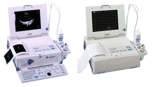 From Fukuda Denshi Co., Ltd.;
'Ultrasound Scanner with built-in Electrocardiograph Realizes Diagnostic Ultrasound Imaging and 12-lead ECG examination with one unit Large 10.4-inch TFT color LCD adopted Cine memory of maximum 64 frames is available Four types of Color Scale Imaging facilitate identification of delicate signals High Frame Rate mode powerfully supports the Cardiovascular region Image data filing in IC card for data management and for easy comparison with past data M-mode continuous recording available with 3-ch ECG data from the cine memory A highly precise and quality printer is integrated Enhanced 12-lead ECG analysis report provided Hybrid and compact design ensures space saving' • 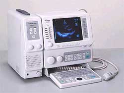 From Fukuda Denshi Co., Ltd.;
'Phased array probes made available for High resolution imaging of full cardiovascular examinations. By adding Linear and Convex probes, the examination areas are extended to Abdominal and OB/GYN applications. B-mode (1, 2 displays), M, and B/M-modes can be individually shown. Optional Doppler unit available for observation of Blood flow information in pulsed and continuous wave Doppler. Portable with IP (Image Processing), Full Keyboard, and track ball are standard.' Result Pages : | Share This Page     Look Ups |
Medical-Ultrasound-Imaging.com
former US-TIP.com
Member of SoftWays' Medical Imaging Group - MR-TIP • Radiology TIP • Medical-Ultrasound-Imaging
Copyright © 2008 - 2025 SoftWays. All rights reserved.
Terms of Use | Privacy Policy | Advertise With Us
former US-TIP.com
Member of SoftWays' Medical Imaging Group - MR-TIP • Radiology TIP • Medical-Ultrasound-Imaging
Copyright © 2008 - 2025 SoftWays. All rights reserved.
Terms of Use | Privacy Policy | Advertise With Us
[last update: 2025-04-05 02:38:00]