Medical Ultrasound Imaging
Wednesday, 8 May 2024
'Color Doppler' Searchterm 'Color Doppler' found in 49 articles 3 terms [ • ] - 46 definitions [• ] Result Pages : • Color Doppler
(CD) Color Doppler is an ultrasound imaging mode, which visualizes the presence, direction and velocity of flowing blood in a wide range of flow conditions. It provides an estimate of the mean velocity of flow within a vessel by color coding the flow and displaying it superimposed on the 2D gray scale image. The flow direction is arbitrarily assigned the color red or blue, indicating flow toward or away from the transducer. Color (colour, Brit.) Doppler ultrasound is capable of evaluating a wider area than other Doppler modes than for example Duplex or power Doppler, and therefore makes it less likely to miss flow abnormalities. It is also easier to interpret. Color flow is not as precise as conventional Doppler and is best used to scan a larger area and then use conventional Doppler for detailed analysis at a site of potential flow abnormality. Adjustments for color Doppler in case of too much color: Adjustments for color Doppler in case of not enough color:
•
increased color gain;
•
decrease color velocity scale;
•
adjust scanning plane and angle to flow;
•
decrease sample box size;
•
evaluation of chosen filter.
See also Color Power Doppler, Autocorrelation, Color Priority, Triplex Exam and Color Saturation. Further Reading: Basics:
News & More:
•
(CDI) Color Doppler imaging depicts the mean frequency shifts of the Doppler signal. Color [colour, Brit.] Doppler imaging is a method for visualizing direction and velocity of movement, such as of blood flow within the cardiac chambers or blood vessels. The flow direction and velocity information gathered by Doppler ultrasonography is color coded onto a gray scale cross-sectional image. The sensitivity of Doppler ultrasound is increased in conjunction with the use of vascular contrast agents. Direction and blood flow velocity are coded as colors and shades: Red - flow coming nearer to the probe. Blue - flow coming away of the probe. See also Bi-directional Illumination, Color Map. Further Reading: News & More:
•
(CDFI) Color [colour, Brit.] Doppler flow imaging is a method based on pulsed ultrasound Doppler technology for visualizing direction and velocity of blood flow within the cardiac chambers or blood vessels.
See also Autocorrelation. •  From SIUI Inc.;
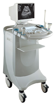
'Dedicated to ultrasound industry, Shantou Institute of Ultrasonic Instruments, Inc. (SIUI) has launched Apogee 3500, the Digital Color Doppler Ultrasound Imaging System. With latest imaging technologies, high-definition image quality and excellent practical functions, the Apogee 3500 offers optimal solutions for clinical ultrasonic examination.' 'The Apogee 3500 is available with many high-density, super broadband and multi-frequency probes, such as convex, micro-convex, linear, vaginal, rectal and phased array probes, which are widely applied for different clinical diagnoses, including abdomen (liver, kidney, gall-bladder, pancreas), gynecology (uterus, ovary), obstetrics (early pregnancy, basic OB, complete OB, multi gestation, fetal echo), cardiology (adult and pediatric cardiology), small parts (thyroid, galactophore, testicles, neonate), peripheral vascular and prostate.'
Device Information and Specification
CONFIGURATION
Normal system, color - gray scale(256)
Linear, convex and phased array
PROBES STANDARD
2.0 MHz ~ 12.0 MHz, broad band, tri-frequency
B-mode (B, 2B, 4B), M-mode, B/M-mode, real-time compound imaging, panoramic imaging, trapezoidal imaging (linear probes), spectrum Doppler (PWD and CWD), color Doppler flow imaging (CDFI), color power angio (CPA), tissue harmonic imaging (THI)
IMAGING OPTIONS
Real-time ZOOM, zoom rate and position selectable
OPTIONAL PACKAGE
H*W*D m
1.29 * 0.52 * 0.75
WEIGHT
110 kg
POWER REQUIREMENT
AC 220V/110V, 50Hz/60Hz
POWER CONSUMPTION
0.6 KVA
•
(CPD) CPD is a type of color Doppler to visualize the presence of detectable blood flow. The flow information is based on the amplitude or strength of echoes received from moving cells and not on frequency shifts. Power Doppler is very sensitive to flowing blood but does not provide velocity or directional information. CPD is less angle dependent than traditional color Doppler, but more sensitive to motion artifacts. Color power angio (CPA) provides better sensitivity to slow flow states. The color maps for CPD are represented by a single continuous color (colour, Brit.). Because CPD does not provide directional information, no aliasing artifact occurs. See also Directional Color Power Doppler. Further Reading: Basics:
Result Pages : |
Medical-Ultrasound-Imaging.com
former US-TIP.com
Member of SoftWays' Medical Imaging Group - MR-TIP • Radiology TIP • Medical-Ultrasound-Imaging
Copyright © 2008 - 2024 SoftWays. All rights reserved.
Terms of Use | Privacy Policy | Advertise With Us
former US-TIP.com
Member of SoftWays' Medical Imaging Group - MR-TIP • Radiology TIP • Medical-Ultrasound-Imaging
Copyright © 2008 - 2024 SoftWays. All rights reserved.
Terms of Use | Privacy Policy | Advertise With Us
[last update: 2023-11-06 01:42:00]


