Medical Ultrasound Imaging
Sunday, 19 May 2024
Info Sheets Out- side     | 'Color Doppler' p3 Searchterm 'Color Doppler' found in 49 articles 3 terms [ • ] - 46 definitions [• ] Result Pages : • 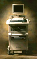 From SIUI Inc.;
'The Apogee 800Plus ultrasound system offers a new level of high quality in image. With broad clinical application, the system delivers ease of use for cardiac and vascular exams.' 'Due to the exceptional image quality, the sensitivity of color Doppler and many other advanced features, the Apogee 800Plus offers complete diagnosis capability for your entire patient population, including abdominal, obstetrics, gynecology, adult cardiology, pediatric cardiology, peripheral vascular, small parts, breast, prostate, transcranial, etc.'
Device Information and Specification
APPLICATIONS
Linear, curved, phased and symmetric phased array
2D, M-mode, Pulsed wave Doppler, Color Power Angio Imaging, Color 2D, Color M-mode, High PRF Doppler
H*W*D m
1.46 * 0.69 * 0.87
WEIGHT
140 kg (without peripherals)
POWER REQUIREMENT
115Vac, (V~), @ 50 to 60 Hz, 1.2kVA
230Vac, (V~), @ 50 to 60 Hz, 1.2kVA POWER CONSUMPTION
800 Watts (with optional OEM'S: 1200 Watts max)
•
An image artifact is any image attribute, which is not present in the original imaged object. An image artifact is sometime the result of an improper operation of the imager, and in other times a consequence of natural processes or properties of the human body. Artifacts in diagnostic ultrasound are a reflection or an echo, which appears on the display and represents the real anatomical structure not correctly. An artifact can be a false, multiple or misleading information introduced by the imaging system or by interaction of ultrasound with the adjacent tissue. Artifacts in ultrasound can be classified as to their source like e.g.:
•
physiologic (motion, different sound velocities, acoustical impedances of tissue);
•
hardware (dimension of the ultrasound beam and the transducer array);
•
Image artifacts can occur in each medical ultrasound. Then an interpretation of the image is complicated and can eliminate the structural information of objects looking for. See also Ultrasound Imaging Procedures. •
The autocorrelation is a mathematical procedure used to quantify periodicity in a Doppler signal and forms the basis of most color Doppler velocity estimators. The autocorrelation multiplies waveforms by successively time-shifted sections of itself.
•
Bi-directional flow is measured in positive and negative directions. See also Bi-directional Illumination, and Color Doppler Imaging. •
Ultrasound at the microbubble resonance frequency can cause bubble rupture at high acoustic power (mechanical index (MI) greater than 0.5). The result is a transient high-amplitude, broadband signal containing all frequencies, not only the harmonics. It will create a strong signal in B-mode or a short-lasting multicolored, mosaic-like effect in color Doppler sonography. Several terms for this typical signal have been used, e.g. induced or stimulated acoustic emission, loss of correlation imaging and sono-scintigraphy. Result Pages : | Share This Page Look Ups |
Medical-Ultrasound-Imaging.com
former US-TIP.com
Member of SoftWays' Medical Imaging Group - MR-TIP • Radiology TIP • Medical-Ultrasound-Imaging
Copyright © 2008 - 2024 SoftWays. All rights reserved.
Terms of Use | Privacy Policy | Advertise With Us
former US-TIP.com
Member of SoftWays' Medical Imaging Group - MR-TIP • Radiology TIP • Medical-Ultrasound-Imaging
Copyright © 2008 - 2024 SoftWays. All rights reserved.
Terms of Use | Privacy Policy | Advertise With Us
[last update: 2023-11-06 01:42:00]