Medical Ultrasound Imaging
Wednesday, 8 May 2024
Info Sheets Out- side     | 'Focal Zone' Searchterm 'Focal Zone' found in 6 articles 1 term [ • ] - 5 definitions [• ] Result Pages : • Focal Zone
The focal zone is the region within the transmitted sound beam in which the beam narrows to its minimum size. The lateral resolution is best within the focal zone of the beam.
•
A zone is a focal region of the ultrasound beam. An ultrasound beam can be directed and focused at a transmit focal zone position. The axial length of the transmit focal zone is a function of the width of the transmit aperture. The field to be imaged is deepened by focusing the transmit energy at progressively deeper points in the body, caused by the beam properties. Typically, multiple zones are used. The main reason for multiple zones is that the transmit energy needs to be greater for points that are deeper in the body, because of the signal's attenuation as it travels into the body. Beam zones:
•
Near zone - the region of a sound beam in which the beam diameter decreases as the distance from the transducer increases (Fresnel zone).
•
Focal zone - the region where the beam diameter is most concentrated giving the greatest degree of focus.
•
Far zone - the region where the beam diameter increases as the distance from the transducer increases (Fraunhofer zone).
The tightest focus and the narrowest beam widths for most conventional transducers are in the mid-field within the zone where the acoustic lens is focused. The ultrasound beam is less well focused and, therefore, wider in the near and far fields which are superficial and deep to the elevation plane focal zone. The beam width is greater in the near and far fields, making lesions in these locations more subject to a partial volume artifact. See also Derated Quantity. •
The acoustic lens is placed at the time the transducer is manufactured and cannot be changed. The acoustic lens is generally focused in the mid field rather than the near or far fields. The exact focal length varies with transducer frequency, but is generally in the range of 4-6 cm for a 5 MHz curved linear probe and 7-9 cm for a 3.5 MHz curved transducer. Placing the elevation plane (z-plane) focal zone of the acoustic lens in the very near or far field would improve the beam width at precisely those depths. However, this would degrade the beam width to a much greater and unacceptable degree at all other depths. There are some chemicals in ultrasound couplants that can degrade the acoustic lens, destroy bonding, or change the acoustic properties of the lens. Problematic chemicals include mineral oil, silicone oil, alcohol, surfactants, and fragrances. Fragrance can affect the transducer's acoustic lens or face material by absorption over time into elastomer and plastic materials, thus changing the material's weight, size, density, and acoustic impedance. Surfactants can degrade the bond between the lens and the piezoelectric elements and contribute to the accelerated degeneration of the lens. See also Retrolenticular Afterglow. Further Reading: Basics:
News & More:
• 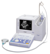 From Fukuda Denshi Co., Ltd.;
'Multi-Purpose Portable Scanner Multi-Frequency Function enables selection of 3 frequencies in each Transducer High-End image Control Function as Zoom, Image Processing, and Variable Focal Zones Built-in Large Track Ball allows ease of use B, B/B, M, and B/M-mode display General Calculation Package for OB, Distance, Area, Volume, and Cardiovascular OB Calculation shown on report page Wide Band, High sensitivity New optional probes' •
Linear array transducer elements are rectangular and arranged in a line. Linear array probes are described by the radius of width in mm. A linear array transducer can have up to 512 elements spaced over 75-120 mm. The beam produced by such a narrow element will diverge rapidly after the wave travels only a few millimeters. The smaller the face of the transducer, the more divergent is the beam. This would result in poor lateral resolution due to beam divergence and low sensitivity due to the small element size. In order to overcome this, adjacent elements are pulsed simultaneously (typically 8 to 16; or more in wide-aperture designs). In a subgroup of x elements, the inner elements pulse delayed with respect to the outer elements. The interference of the x small divergent wavelets produces a focused beam. The delay time determines the depth of focus for the transmitted beam and can be changed during scanning. Linear arrays are usually cheaper than sector scanners but have greater skin contact and therefore make it difficult to reach organs between ribs such as the heart. One-dimensional linear array transducers may have dynamic, electronic focusing providing a narrow ultrasound beam in the image plane. In the z-plane (elevation plane - perpendicular to the image plane) focusing may be provided by an acoustic lens with a fixed focal zone. Rectangular or matrix transducers with unequal rows of transducer elements are two-dimensional (2D), but they are termed 1.5D, because the number of rows is much less than the number of columns. These transducers provide dynamic, electronic focusing even in the z-plane. See also Rectangular Array Transducer. Result Pages : | Share This Page Look Ups |
Medical-Ultrasound-Imaging.com
former US-TIP.com
Member of SoftWays' Medical Imaging Group - MR-TIP • Radiology TIP • Medical-Ultrasound-Imaging
Copyright © 2008 - 2024 SoftWays. All rights reserved.
Terms of Use | Privacy Policy | Advertise With Us
former US-TIP.com
Member of SoftWays' Medical Imaging Group - MR-TIP • Radiology TIP • Medical-Ultrasound-Imaging
Copyright © 2008 - 2024 SoftWays. All rights reserved.
Terms of Use | Privacy Policy | Advertise With Us
[last update: 2023-11-06 01:42:00]