Medical Ultrasound Imaging
Thursday, 9 May 2024
Info Sheets Out- side     | 'Image Quality' p8 Searchterm 'Image Quality' found in 40 articles 1 term [ • ] - 39 definitions [• ] Result Pages : •
A phantom is used to control the imaging performance of ultrasound transducers. The spatial resolution, dead zone, linear fidelity, depth of penetration and image uniformity is important for the image quality. For the axial and lateral resolution, the standard definition is the resolution of objects parallel and perpendicular to the path of the sound beam. Ultrasound pictures created by scans of specially designed ultrasound phantoms can quantify the imaging quality and transducer performance. Phantoms contain one or more materials that simulate a tissue in its interaction with ultrasound. Several phantoms are available specifically for quality control. Transducer characterization consists of a standard pulse echo analysis and insertion loss measurement for each probe. The quality variation from the baseline should be tracked over a period. Further Reading: Basics: •
Ultrasound machines, with their various components and types, have revolutionized the field of medical imaging. These devices enable healthcare professionals to visualize internal structures, assess conditions, and guide interventions with real-time imaging capabilities.
Today, medical ultrasound systems are complex signal processing machines. Assessing the performance of an ultrasound system requires understanding the relationships between the characteristics of the system, such as the point spread function, temporal resolution, and the quality of images. Image quality aspects include the detail resolution, contrast resolution and penetration. Systems with microbubble scanner modification are particularly suitable for contrast enhanced ultrasound.
•
Low-performance systems constitute approximately 20% of the world ultrasound market. These ultrasound machines are characterized by basic black and white imaging and are primarily used for basic OB/GYN applications and fetal development monitoring. They are often purchased by private office practitioners and small hospitals, with a unit cost below $50,000. These scanners commonly come equipped with a transvaginal probe.
•
Mid-performance sonography systems also hold around 20% market share. These machines are basic gray scale imaging, color and spectral Doppler and are used for routine examinations and reporting. They typically utilize a minimum number of scanheads and find applications in radiology, cardiology, and OB/GYN. The cost of these systems ranges between $50,000 and $100,000. Refurbished advanced and high-performance ultrasound machines with fewer optional features can also be found in this price range.
•
High-performance ultrasound systems generally provide high-resolution gray scale imaging, advanced color power and spectral Doppler capabilities. They usually include advanced measurement and analysis software, image review capabilities, and a variety of probes. These high-performance sonography devices have a market share of approximately 40% and cost between $100,000 and $150,000.
•
The remaining 20% of the market consists of premium or advanced performance ultrasound systems, typically sold for over $150,000. Premium performance systems offer high-resolution gray scale imaging, advanced color flow, power Doppler, and spectral Doppler, as well as features like tissue harmonic imaging, image acquisition storage, display and review capabilities, advanced automation, and more. Premium systems are equipped with a wide assortment of transducer scanheads.
In summary, ultrasound machines have diverse performance levels and corresponding price ranges, catering to various medical imaging needs. From low-performance systems with basic imaging capabilities to high-performance and premium systems with advanced features, ultrasound technology continues to advance healthcare imaging capabilities. See also Ultrasound Physics, Handheld Ultrasound, Environmental Protection, Equipment Preparation. Further Reading: Basics:
•
A validation is a test to confirm calibration and accuracy of a measurement, usually by comparing to a known standard such as timed collection. See also Image Quality. •
Conventional, CT and MR imaging technologies are limited in their availability, to depict soft tissue, or to show dynamic activity, like cardiac muscle contractility and blood flow. Easy applicability, real-time sonography and biopsy facilitation are important advantages in veterinarian medicine. Veterinary ultrasound has a very high sensitivity to show the composition of soft tissues, but the low specificity is a disadvantage. High ultrasound system performance includes Doppler techniques, contrast enhanced ultrasound, 3D ultrasound, and tissue harmonic imaging to improve resolution. Technical and physical requirements of veterinary ultrasound are the same as in human ultrasonography. The higher the sound frequency, the better the possible resolution, but the poorer the tissue penetration. Image quality is depended of the ultrasound equipment. For example, a 10 MHz transducer is excellent for imaging of superficial structures; a 3.5 or 5.0 megahertz transducer allows sufficient penetration to see inner structures like the liver or the heart. In addition, the preparation and performing of the examination is similar to that of humans. The sound beam penetrates soft tissue and fat well, but gas and bone impede the ultrasonic power. Fluid filled organs like the bladder are often used as an acoustic window, and an ultrasound gel is used to conduct the sound beam. • 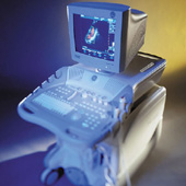 From GE Healthcare.;
(CV Ultrasound) Vivid 3 - 'Vivid family system designed to address customer requests for another level of cardiovascular performance, and offer an even wider range of clinical applications in vascular, pediatrics and the OR. From color flow imaging with super-high frame rates to DICOM connectivity, the new Vivid 3 offers a wealth of technological innovations that enhance image quality, productivity and patient care.' Specifications for this system will be available soon. Result Pages : | Share This Page Look Ups |
Medical-Ultrasound-Imaging.com
former US-TIP.com
Member of SoftWays' Medical Imaging Group - MR-TIP • Radiology TIP • Medical-Ultrasound-Imaging
Copyright © 2008 - 2024 SoftWays. All rights reserved.
Terms of Use | Privacy Policy | Advertise With Us
former US-TIP.com
Member of SoftWays' Medical Imaging Group - MR-TIP • Radiology TIP • Medical-Ultrasound-Imaging
Copyright © 2008 - 2024 SoftWays. All rights reserved.
Terms of Use | Privacy Policy | Advertise With Us
[last update: 2023-11-06 01:42:00]