Medical Ultrasound Imaging
Monday, 20 May 2024
Info Sheets Out- side     | 'Resolution' p5 Searchterm 'Resolution' found in 71 articles 5 terms [ • ] - 66 definitions [• ] Result Pages : •
The prostate is a walnut-shaped gland surrounding the beginning of the urethra in front of the rectum and below the bladder. The prostate can become enlarged (particularly in men over age 50) and develop diseases like prostate cancer or inflammation (prostatitis). A large tumor can be felt by a rectal examination. The most effective way of detecting the early signs of prostate cancer is a combination of a prostate-specific antigen (PSA) blood test and a prostate ultrasound examination. An abnormally high level of PSA can indicate prostate cancer or other prostate diseases such as benign prostatic hypertrophy or prostatitis. The transrectal sonography is an important diagnostic ultrasound procedure in determining whether there is any benign enlargement of the prostate or any abnormal nodules. The imaging is performed with a rectal probe, yielding high resolution. High resolution 3D ultrasound provides reliable and accurate determination of the size and the location of cancer. Additionally, ultrasound elastography is a technique in development to improve the specificity and sensitivity of cancer detection. Ultrasound is also used to detect whether cancerous tissue is still only within the prostate or whether it has begun to spread out and to guide a diagnostic biopsy or ultrasound therapy. See also Brachytherapy, and High Intensity Focused Ultrasound. Further Reading: News & More:
•
(PII) Pulse inversion imaging (also called phase inversion imaging) is a non-linear imaging method specifically made for enhanced detection of microbubble ultrasound contrast agents. In PII, two pulses are sent in rapid succession into the tissue; the second pulse is a mirror image of the first. The resulting echoes are added at reception. Linear scattering of the two pulses will give two echoes which are inverted copies of each other, and these echoes will therefore cancel out when added. Linear scattering dominates in tissues. Echoes from linear scatterers such as tissue cancel, whereas those from gas microbubbles do not. Non-linear scattering of the two pulses will give two echoes which do not cancel out completely due to different bubble response to positive and negative pressures of equal magnitude. The harmonic components add, and the signal intensity difference between non-linear and linear scatterers is therefore increased. The resulting images show high sensitivity to bubbles at the resolution of a conventional image. In harmonic imaging, the frequency range of the transmitted pulse and the received signal should not overlap, but this restriction is less in pulse inversion imaging since the transmit frequencies are not filtered out, but rather subtracted. Broader transmit and receive bandwidths are therefore allowed, giving shorter pulses and improved axial resolution, hence the alternative term wideband harmonic imaging. Many ultrasound machines offer some form of pulse inversion imaging. See also Pulse Inversion Doppler, Narrow Bandwidth, Dead Zone, Ultrasound Phantom. •  From Medison Co.,Ltd.;
'The SONOACE 9900 offers exceptional B/W image clarity by newly developed FINE™ Filter technology, which contributes to edge enhancement and average filtering in real-time. OSIO™(Organ Specified Image Optimization)sets the best diagnosis environment with minimum manual operation and increases patient throughput. Also, color images are improved with better color Doppler performance, high color sensitivity, fine pixel image, and reduced flash artifact across all probes.' 'The SONOACE 9900 PRIME from Medison offers full range of applications specially designed for your practice. With the addition of the new C-Square Processing and PSAD Beamformer, SONOACE 9900 PRIME provides higher contrast resolution and clearer image quality in all application ranges. Based on super clear image resolution and versatile 3D functions, SONOACE 9900 PRIME offers you the true value of future ultrasound technology.' • 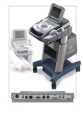 From SonoSite, Inc.;
'Introducing the SonoSite TITAN, a high-resolution modular ultrasound solution. TITAN provides health care professionals with state of the art high-resolution imaging wherever needed for responsive, rapid patient care. The TITAN system's modular design includes three major benefits: Stationary and mobile usage. Point-of-care connectivity. Flexible, cost-effective paths that enable users to acquire new components and software applications.' •
Conventional, CT and MR imaging technologies are limited in their availability, to depict soft tissue, or to show dynamic activity, like cardiac muscle contractility and blood flow. Easy applicability, real-time sonography and biopsy facilitation are important advantages in veterinarian medicine. Veterinary ultrasound has a very high sensitivity to show the composition of soft tissues, but the low specificity is a disadvantage. High ultrasound system performance includes Doppler techniques, contrast enhanced ultrasound, 3D ultrasound, and tissue harmonic imaging to improve resolution. Technical and physical requirements of veterinary ultrasound are the same as in human ultrasonography. The higher the sound frequency, the better the possible resolution, but the poorer the tissue penetration. Image quality is depended of the ultrasound equipment. For example, a 10 MHz transducer is excellent for imaging of superficial structures; a 3.5 or 5.0 megahertz transducer allows sufficient penetration to see inner structures like the liver or the heart. In addition, the preparation and performing of the examination is similar to that of humans. The sound beam penetrates soft tissue and fat well, but gas and bone impede the ultrasonic power. Fluid filled organs like the bladder are often used as an acoustic window, and an ultrasound gel is used to conduct the sound beam. Result Pages : | Share This Page Look Ups |
Medical-Ultrasound-Imaging.com
former US-TIP.com
Member of SoftWays' Medical Imaging Group - MR-TIP • Radiology TIP • Medical-Ultrasound-Imaging
Copyright © 2008 - 2024 SoftWays. All rights reserved.
Terms of Use | Privacy Policy | Advertise With Us
former US-TIP.com
Member of SoftWays' Medical Imaging Group - MR-TIP • Radiology TIP • Medical-Ultrasound-Imaging
Copyright © 2008 - 2024 SoftWays. All rights reserved.
Terms of Use | Privacy Policy | Advertise With Us
[last update: 2023-11-06 01:42:00]