Medical Ultrasound Imaging
Sunday, 19 May 2024
Info Sheets Out- side     | 'Sound' p20 Searchterm 'Sound' found in 496 articles 64 terms [ • ] - 432 definitions [• ] Result Pages : •
(TRUS) Transrectal sonography (also called transrectal ultrasonography, transrectal echography (TRE), endorectal ultrasound (ERUS or EUS)) is an ultrasound procedure used to examine the prostate gland, the rectum or bladder. A small, lubricated transducer placed into the rectum releases sound waves, which create echoes as they enter the region of interest. A computer creates a picture called a sonogram. TRUS is commonly used for guidance during a prostate needle biopsy and may be used to deliver brachytherapy and monitor cancer treatment. Transrectal ultrasonography detects enlargement, tumors and other abnormalities of the prostate, rectal polyps, rectal cancer, perianal infection, and sphincter muscle injuries. TRUS is also performed on male patients with infertility to view the prostate and surrounding structures and on patients with suspected bladder conditions or disease to view the bladder. See also Transurethral Sonography, Endoscopic Ultrasound, Pelvic Ultrasound, Rectal Probe, Biplane Probe, Endocavitary Echography and High Intensity Focused Ultrasound. Further Reading: News & More:
•  From Verathon Inc.;
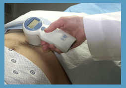
'The BladderScan® BVI 6100 is a handheld, noninvasive ultrasound instrument that measures bladder volume. It is easy to operate, so any staff member can scan patients quickly and accurately. To view ultrasound images from exams and print BVI 6100 exam results for patient records and reimbursement, just log on to ScanPoint®, Verathon Inc.'s (formerly Diagnostic Ultrasound Corp.) innovative online service. The BVI 6100 makes it easier to provide the best care for your patients.' See also Urologic Ultrasound, Mirror Artifact, Pelvic Ultrasound, Transrectal Sonography and Ultrasonography. •
Brachytherapy is a radiation therapy in which radioactive material (radioisotopes) sealed in needles, seeds or wires is placed directly into or near a tumor. Brachytherapy uses ultrasound imaging to visualize the needles for accurate placement of the small seeds or pellets (capsules) directly into e.g., the prostate. Ultrasound imaging allows accurate planning, placement and implantation of the radiation sources. Implantation of the seeds is a minimally invasive procedure. Radioactive seeds are inserted through the perineum skin (the area between the scrotum and the anus) into the prostate gland. With correct planning, the surgeon can implant the radiation sources for maximum benefits to effective cancer treatment. See also EchoSeed™, Prostate Ultrasound, Thermotherapy, High Intensity Focused Ultrasound, Urologic Ultrasound, Transurethral Sonography. Further Reading: News & More:
•
Cavitation is any activity of highly compressible transient or stable microbubbles of gas and/or vapour, generated by ultrasonic power in the propagation medium. Cavitation can be described as inertial or non-inertial. Inertial cavitation has the most potential to damage tissue and occurs when a gas-filled cavity grows, during pressure rarefaction of the ultrasound pulse, and contracts, during the compression phase. Collapses of bubbles can generate local high temperatures and pressures. Transient cavitation can cause tissue damage.
The threshold for cavitation is high and does not occur at current levels of diagnostic ultrasound. The introduction of contrast agents leads to the formation of microbubbles that potentially provide gas nuclei for cavitation. The use of contrast agents can lower the threshold at which cavitation occurs. Types of cavitation:
•
Stable cavitation - steady microbubble oscillation due to the passage of a sound wave.
•
Cross-section scattering is a measure of the scattering strength of a point scatterer. The scattering strength is dependent on the size of the scatterer, the density and compressibility of the scatterer and the surrounding medium, and the ultrasound wavelength. If a transducer emits ultrasound with a total acoustic power of P, and the power is assumed to be uniform distributed over the US beam cross-sectional area, then the ultrasound intensity at a certain range, is defined by: I = P/A where I is the intensity, and A is the cross-sectional beam area at that range. A point scatterer located in the ultrasound beam at this range, will scatter the ultrasound with a total acoustic power of Ps, defined by: Ps = I s where s is the scattering cross-section of the point scatterer. Result Pages : | Share This Page Look Ups |
Medical-Ultrasound-Imaging.com
former US-TIP.com
Member of SoftWays' Medical Imaging Group - MR-TIP • Radiology TIP • Medical-Ultrasound-Imaging
Copyright © 2008 - 2024 SoftWays. All rights reserved.
Terms of Use | Privacy Policy | Advertise With Us
former US-TIP.com
Member of SoftWays' Medical Imaging Group - MR-TIP • Radiology TIP • Medical-Ultrasound-Imaging
Copyright © 2008 - 2024 SoftWays. All rights reserved.
Terms of Use | Privacy Policy | Advertise With Us
[last update: 2023-11-06 01:42:00]