Medical Ultrasound Imaging
Sunday, 19 May 2024
Info Sheets Out- side     | 'Thyroid Ultrasound' p3 Searchterm 'Thyroid Ultrasound' found in 13 articles 1 term [ • ] - 1 definition [• ] - 11 booleans [• ]Result Pages : • 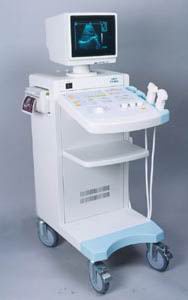 From SIUI Inc.;
'The CTS-310B is designed for the diagnosis of liver, gall, kidney, pancreas, thyroid, breast, uterus, bladder, ovary, etc. It is a versatile ultrasound scanner with both linear array scanning and convex scanning.' Features: 'Powerful image processing circuit & High quality image A wide range of probes for selection Cineloop Probe frequency conversation option Computer image communication Various measuring function Backlit keyboard'
Device Information and Specification
APPLICATIONS
See description above
CONFIGURATION
Normal system, 10' high resolution monitor, dual probe connector
Linear and convex
PROBES STANDARD
2.5MHz to 10.0MHz, linear and convex, broad band, trifrequency
IMAGING OPTIONS
Multi zoom rate and depth shift
OPTIONAL PACKAGE
POWER REQUIREMENT
AC 220V/110V, 50Hz/60Hz
POWER CONSUMPTION
0.1 KVA
•  From SIUI Inc.;
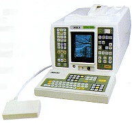
'The SIUI CTS-200 is one of few 'linear only' units to be developed with a digital scan converter to process all incoming signals. With a digital processor, not only is the incoming signal processed faster but less noise is introduced into the signal path. The images are cleaner, crisper than you have experienced, even in machines at many times the price.' 'CTS-200 is suitable to a wide range of examination of liver, gallbladder, kidney, spleen, pancreas, thyroid gland, breast, uterus, urinary bladder, OB/GYN, etc. It is a portable ultrasound scanner of high performance.'
Device Information and Specification
APPLICATIONS
See description above
CONFIGURATION
Portable, gray scale(256)
Linear
PROBES STANDARD
1 * 3.5MHz Linear Probe EZU-PL21
PROBES FREQUENCY
3.5MHz, 7.5MHz
IMAGING OPTIONS
*1, *1.5, *2 as well as depth shift
OPTIONAL PACKAGE
7.5MHz Linear Probe EZU-PL23; 7.5MHz Transrectal Probe EUP-U23; 3.5MHz Biopsy Probe EUP-B11A; Trolley/Mobile Cart; ...
DATA PROCESSING
Pre-processing, correlation-processing, interpolation
POWER REQUIREMENT
AC 220V/110V, 50Hz/60Hz
POWER CONSUMPTION
0.05 KVA
•
Ultrasound elastography is a specialized imaging technique that provides information about tissue elasticity or stiffness. It is used to assess the mechanical properties of tissues, helping to differentiate between normal and abnormal tissue conditions.
The basic principle behind ultrasound elastography involves the application of mechanical stress to the tissue and measuring its resulting deformation. This is typically achieved by using either external compression or shear waves generated by the ultrasound transducer. There are two main types of ultrasound elastography: •
Strain Elastography: In strain elastography, the tissue is mechanically compressed using the ultrasound transducer, causing deformation. The transducer then captures images before and after compression, and the software analyzes the displacement or strain between these images. Softer tissues tend to deform more than stiffer tissues, and this information is used to generate a color-coded map or elastogram, where softer areas appear in different colors compared to stiffer regions.
•
Shear Wave Elastography: Shear wave elastography involves the generation of shear waves within the tissue using focused ultrasound beams. These shear waves propagate through the tissue, and their velocity is measured using the ultrasound transducer. The speed of shear wave propagation is directly related to tissue stiffness: stiffer tissues transmit shear waves faster than softer tissues. By calculating the shear wave velocity, an elastogram is generated, providing a quantitative assessment of tissue stiffness.
Both strain elastography and shear wave elastography offer valuable insights into tissue characteristics and can assist in the diagnosis and characterization of various conditions. In clinical practice, ultrasound elastography is particularly useful for evaluating liver fibrosis, breast lesions, thyroid nodules, prostate abnormalities, and musculoskeletal conditions. By providing additional information about tissue stiffness, ultrasound elastography enhances the diagnostic capabilities of traditional ultrasound imaging. It allows for non-invasive assessment, improves the accuracy of tissue characterization, and aids in treatment planning and monitoring of various medical conditions. See also Ultrasound Accessories and Supplies, Sonographer and Ultrasound Technology. Result Pages : | Share This Page Look Ups |
Medical-Ultrasound-Imaging.com
former US-TIP.com
Member of SoftWays' Medical Imaging Group - MR-TIP • Radiology TIP • Medical-Ultrasound-Imaging
Copyright © 2008 - 2024 SoftWays. All rights reserved.
Terms of Use | Privacy Policy | Advertise With Us
former US-TIP.com
Member of SoftWays' Medical Imaging Group - MR-TIP • Radiology TIP • Medical-Ultrasound-Imaging
Copyright © 2008 - 2024 SoftWays. All rights reserved.
Terms of Use | Privacy Policy | Advertise With Us
[last update: 2023-11-06 01:42:00]