Medical Ultrasound Imaging
Friday, 3 May 2024
Info Sheets Out- side     | 'View' Searchterm 'View' found in 43 articles 1 term [ • ] - 42 definitions [• ] Result Pages : •
(FOV) The field of view is the plane or area depicted by the ultrasound transducer.
•
(AUS) Abdominal ultrasound, also known as abdominal sonography, is a medical imaging technique that focuses on the visualization and assessment of the abdominal organs. While 'abdominal ultrasound' is the commonly used term, there are alternative terms that can be used to refer to this imaging modality: (TAE) transabdominal echography, abdominal ultrasonography, sonogram, FAST (Focused Assessment with Sonography for Trauma). Abdominal ultrasound imaging is an invaluable clinical tool for identifying the underlying cause of abdominal pain. An abdominal ultrasound examination encompasses a comprehensive evaluation of the liver, gallbladder, biliary tree, pancreas, spleen, kidneys, and abdominal blood vessels. It is a cost-effective, safe, and non-invasive medical imaging modality that is typically utilized as the initial diagnostic investigation. Advanced ultrasound techniques, such as high-resolution ultrasound, endoscopic ultrasound, and contrast-enhanced Doppler, further enhance the detection of small lesions and provide detailed information for precise diagnosis. To prepare for an abdominal ultrasound, it is recommended to have nothing to eat or drink for at least 8 hours, starting from midnight the night before the examination. Indications:
•
Abdominal pain
•
Gallbladder or kidneys stones
•
Inflammation
•
Detection of cancer and metastasis
FAST (Focused Assessment with Sonography for Trauma) is a rapid diagnostic test used for trauma patients. It sequentially evaluates the presence of free fluid in the pericardium (hemopericardium) and in four specific views of the abdomen. These views include the right upper quadrant (RUQ), left upper quadrant (LUQ), subcostal, and suprapubic views. They aid in identifying hemoperitoneum in patients with potential truncal injuries. The space between the liver and the right kidney (RUQ), known as Morison's pouch, is a location where intraperitoneal fluid can accumulate. Emergency abdominal ultrasonography is indicated in cases of suspected aortic aneurysm, appendicitis, biliary and renal colic, as well as blunt or penetrating abdominal trauma. It plays a crucial role in the timely assessment and management of these conditions, providing critical information to guide appropriate treatment decisions. See also Handheld Ultrasound, Pelvic Ultrasound, Pregnancy Ultrasound, Prostate Ultrasound, Interventional Ultrasound and Pediatric Ultrasound. Further Reading: Basics: News & More:
•
If available, some graphic aids can be helpful to show image orientations. 1) A graphic icon of the labeled primary axes (A, L, H) with relative lengths given by direction sines and orientation as if viewed from the normal to the image plane can help orient the viewer, both to identify image plane orientation and to indicate possible in plane rotation. 2) Ingraphic prescription of obliques from other images, a sample original image with an overlaid line or set of lines indicating the intersection of the original and oblique image planes can help orient the viewer.
•
•
In all cases the scanning surface is assigned to the top of the image. The orientation of single oblique slices can be specified by rotating a slice in one of the basic orientations toward one of the other two basic orthogonal planes about an axis defined by the intersection of the 2 planes. See also Histogram. • 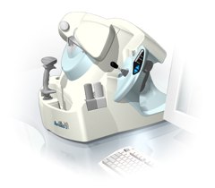 From Ultralink LLC;
'Artemis is a very high frequency (VHF) ultrasound eye scanner. In use, the patient leans forward placing their head onto an adjustable headrest. The headrest's unique design permits the patient to pull away quickly from the scanner if desired. An eyecup filled with a saline-based interface fluid couples the ultrasound signal to the eye, while a precision mechanism moves the transducer past the front of the eye. During the accurately controlled arc motion of the transducer, which lasts less than one second, many thousands of ultrasound samples are digitized. Following a scan, signal analysis is performed on a PC-compatible microcomputer, and the data are available for immediate viewing on an LCD monitor or disk storage. Artemis is very flexible; many adjustments to the scanning parameters are possible to customize the scan to your clinical needs. Functions are provided for centering the scan about the optical axis of the eye. The starting location of the scans as well as the extent can be varied as desired, to view image planes through the eye at different angles.' See also Ultrasound Biomicroscopy, A-Mode and A-Scan. Further Reading: News & More:
•
Convex transducers are today standard on every new scanner. A convex surface allows the scanning of a larger area with a smaller array. The method of focusing and beam sweeping of a convex or curvilinear / curved array is similar to a linear array transducer, except of the shape of the probe and the sector format of the created image. The better fit to the body, caused by the curved shape with smaller convex contact surface, and the wider field of view further from the transducer face are advantages in abdominal ultrasound. However, also a convex array is often too large to image the heart when probing between the ribs. Caused by combining a large field of view with smallest array size, phased array transducers are the best choice in cardiac ultrasound. See also Curved Transducer. Result Pages : | Share This Page Look Ups |
Medical-Ultrasound-Imaging.com
former US-TIP.com
Member of SoftWays' Medical Imaging Group - MR-TIP • Radiology TIP • Medical-Ultrasound-Imaging
Copyright © 2008 - 2024 SoftWays. All rights reserved.
Terms of Use | Privacy Policy | Advertise With Us
former US-TIP.com
Member of SoftWays' Medical Imaging Group - MR-TIP • Radiology TIP • Medical-Ultrasound-Imaging
Copyright © 2008 - 2024 SoftWays. All rights reserved.
Terms of Use | Privacy Policy | Advertise With Us
[last update: 2023-11-06 01:42:00]