Medical Ultrasound Imaging
Sunday, 19 May 2024
Info Sheets Out- side     | '2D Ultrasound' p2 Searchterm '2D Ultrasound' found in 20 articles 1 term [ • ] - 5 definitions [• ] - 14 booleans [• ]Result Pages : •
Ultrasound technology with its advancements is vital for delivering high-quality patient care. Innovations including high-frequency ultrasound, 3D//4D imaging, contrast enhanced ultrasound, elastography, and point-of-care ultrasound, have expanded the capabilities of ultrasound imaging and improved diagnostic accuracy. B-Mode imaging, also known as brightness mode, is the fundamental technique in ultrasound imaging. It produces two-dimensional images based on the echoes received from tissues and organs. Understanding the principles of B-Mode imaging, such as gain adjustment, depth control, and image optimization, is crucial for obtaining diagnostically valuable images. M-Mode imaging, on the other hand, allows for the visualization of motion over time, enabling assessment of cardiac structures and function, as well as fetal heart rate. High-frequency ultrasound refers to the use of ultrasound waves with frequencies greater than 10 MHz. This technology enables improved resolution, allowing for detailed imaging of superficial structures like skin, tendons, and small organs. High-frequency ultrasound has found applications in dermatology, ophthalmology, and musculoskeletal imaging. Traditional 2D ultrasound has been augmented by the advent of 3D ultrasound technology. By acquiring multiple 2D images from different angles, this technique construct a volumetric representation of the imaged area. The addition of 4D ultrasound in real-time motion adds further value by capturing dynamic processes. Doppler imaging employs the Doppler effect to evaluate blood flow within vessels and assess hemodynamics. Color Doppler assigns color to different blood flow velocities, providing a visual representation of blood flow direction and speed. Spectral Doppler displays blood flow velocities as a waveform, allowing for detailed analysis of flow patterns, resistance, and stenosis. Contrast enhanced ultrasound employs microbubble contrast agents to enhance the visualization of blood flow and tissue perfusion. By injecting these agents intravenously, sonographers can differentiate between vascular structures and lesions. Elastography is a technique that measures tissue elasticity or stiffness. It assists in differentiating between normal and abnormal tissues, aiding in the diagnosis of various conditions such as liver fibrosis, breast lesions, and thyroid nodules. Fusion imaging combines ultrasound with other imaging modalities, such as computed tomography (CT), magnetic resonance imaging (MRI), or positron emission tomography (PET). By overlaying or merging ultrasound images with those obtained from other modalities, the user can precisely locate and characterize abnormalities, guide interventions, and improve diagnostic accuracy. Fusion imaging has proven particularly useful in areas such as interventional radiology, oncology, and urology. See also Equipment Preparation, Environmental Protection, Handheld Ultrasound, Portable Ultrasound and Ultrasound Accessories and Supplies. • 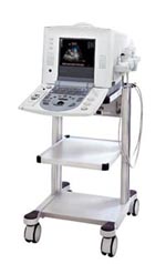 From Kontron Medical SAS;
'Vetson is an Ultrasound System designed for veterinary applications. From 2D to Color Flow Mapping (CFM), Vetson Pro and Vetson Color are the best tools you can find on the veterinary market for quick and confident diagnosis and easy operation.' Specifications for this system will be available soon. • 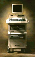 From SIUI Inc.;
'The Apogee 800Plus ultrasound system offers a new level of high quality in image. With broad clinical application, the system delivers ease of use for cardiac and vascular exams.' 'Due to the exceptional image quality, the sensitivity of color Doppler and many other advanced features, the Apogee 800Plus offers complete diagnosis capability for your entire patient population, including abdominal, obstetrics, gynecology, adult cardiology, pediatric cardiology, peripheral vascular, small parts, breast, prostate, transcranial, etc.'
Device Information and Specification
APPLICATIONS
Linear, curved, phased and symmetric phased array
2D, M-mode, Pulsed wave Doppler, Color Power Angio Imaging, Color 2D, Color M-mode, High PRF Doppler
H*W*D m
1.46 * 0.69 * 0.87
WEIGHT
140 kg (without peripherals)
POWER REQUIREMENT
115Vac, (V~), @ 50 to 60 Hz, 1.2kVA
230Vac, (V~), @ 50 to 60 Hz, 1.2kVA POWER CONSUMPTION
800 Watts (with optional OEM'S: 1200 Watts max)
•
A C-scan ultrasound can be displayed in 2D or 3D ultrasound technique. C-scan systems can generate images which are parallel to the surface of the skin (coronal). 2D plane images, usually in gray scale, are recordable at different depths, maintaining high quality subsurface information.
Further Reading: Basics:
News & More:
•
Echo ranging is the ultrasound relationship between transit time and reflector depth expressed as: t = 2d//c Result Pages : | Share This Page Look Ups |
Medical-Ultrasound-Imaging.com
former US-TIP.com
Member of SoftWays' Medical Imaging Group - MR-TIP • Radiology TIP • Medical-Ultrasound-Imaging
Copyright © 2008 - 2024 SoftWays. All rights reserved.
Terms of Use | Privacy Policy | Advertise With Us
former US-TIP.com
Member of SoftWays' Medical Imaging Group - MR-TIP • Radiology TIP • Medical-Ultrasound-Imaging
Copyright © 2008 - 2024 SoftWays. All rights reserved.
Terms of Use | Privacy Policy | Advertise With Us
[last update: 2023-11-06 01:42:00]