Medical Ultrasound Imaging
Sunday, 19 May 2024
'Ultrasound Technology' p4 Searchterm 'Ultrasound Technology' found in 36 articles 1 term [ • ] - 18 definitions [• ] - 17 booleans [• ]Result Pages : •
Ultrasound elastography is a specialized imaging technique that provides information about tissue elasticity or stiffness. It is used to assess the mechanical properties of tissues, helping to differentiate between normal and abnormal tissue conditions.
The basic principle behind ultrasound elastography involves the application of mechanical stress to the tissue and measuring its resulting deformation. This is typically achieved by using either external compression or shear waves generated by the ultrasound transducer. There are two main types of ultrasound elastography: •
Strain Elastography: In strain elastography, the tissue is mechanically compressed using the ultrasound transducer, causing deformation. The transducer then captures images before and after compression, and the software analyzes the displacement or strain between these images. Softer tissues tend to deform more than stiffer tissues, and this information is used to generate a color-coded map or elastogram, where softer areas appear in different colors compared to stiffer regions.
•
Shear Wave Elastography: Shear wave elastography involves the generation of shear waves within the tissue using focused ultrasound beams. These shear waves propagate through the tissue, and their velocity is measured using the ultrasound transducer. The speed of shear wave propagation is directly related to tissue stiffness: stiffer tissues transmit shear waves faster than softer tissues. By calculating the shear wave velocity, an elastogram is generated, providing a quantitative assessment of tissue stiffness.
Both strain elastography and shear wave elastography offer valuable insights into tissue characteristics and can assist in the diagnosis and characterization of various conditions. In clinical practice, ultrasound elastography is particularly useful for evaluating liver fibrosis, breast lesions, thyroid nodules, prostate abnormalities, and musculoskeletal conditions. By providing additional information about tissue stiffness, ultrasound elastography enhances the diagnostic capabilities of traditional ultrasound imaging. It allows for non-invasive assessment, improves the accuracy of tissue characterization, and aids in treatment planning and monitoring of various medical conditions. See also Ultrasound Accessories and Supplies, Sonographer and Ultrasound Technology. •
(US) Also called echography, sonography, ultrasonography, echotomography, ultrasonic tomography. Diagnostic imaging plays a vital role in modern healthcare, allowing medical professionals to visualize internal structures of the body and assist in the diagnosis and treatment of various conditions. Two terms that are commonly used interchangeably but possess distinct meanings in the field of medical imaging are 'ultrasound' and 'sonography.' Ultrasound is the imaging technique that utilizes sound waves to create real-time images, while sonography encompasses the entire process of performing ultrasound examinations and interpreting the obtained images. Ultrasonography is a synonymous term for sonography, emphasizing the use of ultrasound technology in diagnostic imaging. A sonogram, on the other hand, refers to the resulting image produced during an ultrasound examination. Ultrasonic waves, generated by a quartz crystal, cause mechanical perturbation of an elastic medium, resulting in rarefaction and compression of the medium particles. These waves are reflected at the interfaces between different tissues due to differences in their mechanical properties. The transmission and reflection of these high-frequency waves are displayed with different types of ultrasound modes. By utilizing the speed of wave propagation in tissues, the time of reflection information can be converted into distance of reflection information. The use of higher frequencies in medical ultrasound imaging yields better image resolution. However, higher frequencies also lead to increased absorption of the sound beam by the medium, limiting its penetration depth. For instance, higher frequencies (e.g., 7.5 MHz) are employed to provide detailed imaging of superficial organs like the thyroid gland and breast, while lower frequencies (e.g., 3.5 MHz) are used for abdominal examinations. Ultrasound in medical imaging offers several advantages including:
•
noninvasiveness;
•
safety with no potential risks;
•
widespread availability and relatively low cost.
Diagnostic ultrasound imaging is generally considered safe, with no adverse effects. As medical ultrasound is extensively used in pregnancy and pediatric imaging, it is crucial for practitioners to ensure its safe usage. Ultrasound can cause mechanical and thermal effects in tissue, which are amplified with increased output power. Consequently, guidelines for the safe use of ultrasound have been issued to address the growing use of color flow imaging, pulsed spectral Doppler, and higher demands on B-mode imaging. Furthermore, recent ultrasound safety regulations have shifted more responsibility to the operator to ensure the safe use of ultrasound. See also Skinline, Pregnancy Ultrasound, Obstetric and Gynecologic Ultrasound, Musculoskeletal and Joint Ultrasound, Ultrasound Elastography and Prostate Ultrasound. Further Reading: Basics: News & More:
•
Ultrasound machines, with their various components and types, have revolutionized the field of medical imaging. These devices enable healthcare professionals to visualize internal structures, assess conditions, and guide interventions with real-time imaging capabilities.
Today, medical ultrasound systems are complex signal processing machines. Assessing the performance of an ultrasound system requires understanding the relationships between the characteristics of the system, such as the point spread function, temporal resolution, and the quality of images. Image quality aspects include the detail resolution, contrast resolution and penetration. Systems with microbubble scanner modification are particularly suitable for contrast enhanced ultrasound.
•
Low-performance systems constitute approximately 20% of the world ultrasound market. These ultrasound machines are characterized by basic black and white imaging and are primarily used for basic OB/GYN applications and fetal development monitoring. They are often purchased by private office practitioners and small hospitals, with a unit cost below $50,000. These scanners commonly come equipped with a transvaginal probe.
•
Mid-performance sonography systems also hold around 20% market share. These machines are basic gray scale imaging, color and spectral Doppler and are used for routine examinations and reporting. They typically utilize a minimum number of scanheads and find applications in radiology, cardiology, and OB/GYN. The cost of these systems ranges between $50,000 and $100,000. Refurbished advanced and high-performance ultrasound machines with fewer optional features can also be found in this price range.
•
High-performance ultrasound systems generally provide high-resolution gray scale imaging, advanced color power and spectral Doppler capabilities. They usually include advanced measurement and analysis software, image review capabilities, and a variety of probes. These high-performance sonography devices have a market share of approximately 40% and cost between $100,000 and $150,000.
•
The remaining 20% of the market consists of premium or advanced performance ultrasound systems, typically sold for over $150,000. Premium performance systems offer high-resolution gray scale imaging, advanced color flow, power Doppler, and spectral Doppler, as well as features like tissue harmonic imaging, image acquisition storage, display and review capabilities, advanced automation, and more. Premium systems are equipped with a wide assortment of transducer scanheads.
In summary, ultrasound machines have diverse performance levels and corresponding price ranges, catering to various medical imaging needs. From low-performance systems with basic imaging capabilities to high-performance and premium systems with advanced features, ultrasound technology continues to advance healthcare imaging capabilities. See also Ultrasound Physics, Handheld Ultrasound, Environmental Protection, Equipment Preparation. Further Reading: Basics:
• Verathon Inc. formerly 'Diagnostic Ultrasound' was founded in 1984 by Gerald McMorrow. His vision was to use emerging ultrasound technology to develop noninvasive, easy to use medical devices that would fill unmet needs in health care. In the beginning, the company was no more than one man working alone in his basement. Today, the company has 150 employees and has sold more than 100 million dollars worth of its products worldwide. Ultrasound Systems:
Contact Information
ONLINE
CONTACT
•  From ESAOTE S.p.A.;
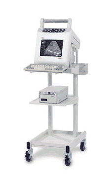
'The wide range of OB/GYN and general ultrasound applications defines the Falco as an all-around ultrasound system, perfectly suited for health care practitioners worldwide. Although it is a compact and portable system, the Falco is equipped with the latest in imaging software, including Esaote's CRII Technology, Total Image Focus and Fine Line Processing.' Result Pages : |
Medical-Ultrasound-Imaging.com
former US-TIP.com
Member of SoftWays' Medical Imaging Group - MR-TIP • Radiology TIP • Medical-Ultrasound-Imaging
Copyright © 2008 - 2024 SoftWays. All rights reserved.
Terms of Use | Privacy Policy | Advertise With Us
former US-TIP.com
Member of SoftWays' Medical Imaging Group - MR-TIP • Radiology TIP • Medical-Ultrasound-Imaging
Copyright © 2008 - 2024 SoftWays. All rights reserved.
Terms of Use | Privacy Policy | Advertise With Us
[last update: 2023-11-06 01:42:00]


