Medical Ultrasound Imaging
Sunday, 10 November 2024
'Echo' p17 Searchterm 'Echo' found in 160 articles 28 terms [ • ] - 132 definitions [• ] Result Pages : •  From Siemens Medical Systems;
We see a way to review 100% of your cardiovascular ultrasound exams and reports anywhere, anytime. The Cypress™ system is a highly miniaturized, all digital, phased array echocardiography system that provides complete studies and outstanding images - even on the most technically difficult patients. Specifications for this system will be available soon. •  From ALOKA Co., Ltd.;
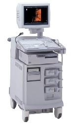
'The ProSound SSD-4000 utilizes the most advanced acoustic technologies available today, and its multidisciplinary technology architecture enables it to offer great versatility and flexibility over a wide range of clinical applications. With its new-generation, front-end technology including a 12-bit A/D converter, the ProSound SSD-4000 offers superior contrast resolution−especially when compared to 10-bit systems.'
Device Information and Specification
APPLICATIONS
CONFIGURATION
Compact, portable, dual dynamic display
Wide-band super high-density (W-SHD) transducers
OPTIONAL PACKAGE
Volume Mode
STORAGE, CONNECTIVITY, OS
Data Management Subsystem (iDMS), DICOM-Worklist
DATA PROCESSING
•  From ALOKA Co., Ltd.;
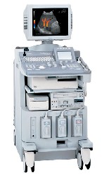
'A Platform for Pure Harmonic Detection The SSD-5000 is the second platform in our ProSound™ line of superior ultrasound systems. The Pure Harmonic Detection technology provides distortion-free fundamental frequency transmission, offering optimum Harmonic Echo™ image quality. This technology is especially effective for obese patients and in a variety of technically difficult scanning conditions.' •
(AUS) Abdominal ultrasound, also known as abdominal sonography, is a medical imaging technique that focuses on the visualization and assessment of the abdominal organs. While 'abdominal ultrasound' is the commonly used term, there are alternative terms that can be used to refer to this imaging modality: (TAE) transabdominal echography, abdominal ultrasonography, sonogram, FAST (Focused Assessment with Sonography for Trauma). Abdominal ultrasound imaging is an invaluable clinical tool for identifying the underlying cause of abdominal pain. An abdominal ultrasound examination encompasses a comprehensive evaluation of the liver, gallbladder, biliary tree, pancreas, spleen, kidneys, and abdominal blood vessels. It is a cost-effective, safe, and non-invasive medical imaging modality that is typically utilized as the initial diagnostic investigation. Advanced ultrasound techniques, such as high-resolution ultrasound, endoscopic ultrasound, and contrast-enhanced Doppler, further enhance the detection of small lesions and provide detailed information for precise diagnosis. To prepare for an abdominal ultrasound, it is recommended to have nothing to eat or drink for at least 8 hours, starting from midnight the night before the examination. Indications:
•
Abdominal pain
•
Gallbladder or kidneys stones
•
Inflammation
•
Detection of cancer and metastasis
FAST (Focused Assessment with Sonography for Trauma) is a rapid diagnostic test used for trauma patients. It sequentially evaluates the presence of free fluid in the pericardium (hemopericardium) and in four specific views of the abdomen. These views include the right upper quadrant (RUQ), left upper quadrant (LUQ), subcostal, and suprapubic views. They aid in identifying hemoperitoneum in patients with potential truncal injuries. The space between the liver and the right kidney (RUQ), known as Morison's pouch, is a location where intraperitoneal fluid can accumulate. Emergency abdominal ultrasonography is indicated in cases of suspected aortic aneurysm, appendicitis, biliary and renal colic, as well as blunt or penetrating abdominal trauma. It plays a crucial role in the timely assessment and management of these conditions, providing critical information to guide appropriate treatment decisions. See also Handheld Ultrasound, Pelvic Ultrasound, Pregnancy Ultrasound, Prostate Ultrasound, Interventional Ultrasound and Pediatric Ultrasound. Further Reading: Basics: News & More:
•
Echoes of deep lying structures within the body do not always come from the latest emitted sound pulse and can produce an aliasing artifact. Aliasing lowers the frequency components when the pulse repetition frequency is less than 2 times the highest frequency of a Doppler signal. This artifact can be problematical at Spectral or Color Doppler examinations. Aliasing of the data displayed in pulsed wave technology is utilized as a benefit in determining transitions from laminar to turbulent flow. See also Ultrasound Imaging Modes. Result Pages : |
Medical-Ultrasound-Imaging.com
former US-TIP.com
Member of SoftWays' Medical Imaging Group - MR-TIP • Radiology TIP • Medical-Ultrasound-Imaging
Copyright © 2008 - 2024 SoftWays. All rights reserved.
Terms of Use | Privacy Policy | Advertise With Us
former US-TIP.com
Member of SoftWays' Medical Imaging Group - MR-TIP • Radiology TIP • Medical-Ultrasound-Imaging
Copyright © 2008 - 2024 SoftWays. All rights reserved.
Terms of Use | Privacy Policy | Advertise With Us
[last update: 2023-11-06 01:42:00]


